
87 Primary Myelofibrosis (PMF)
Michelle To and Valentin Villatoro
-

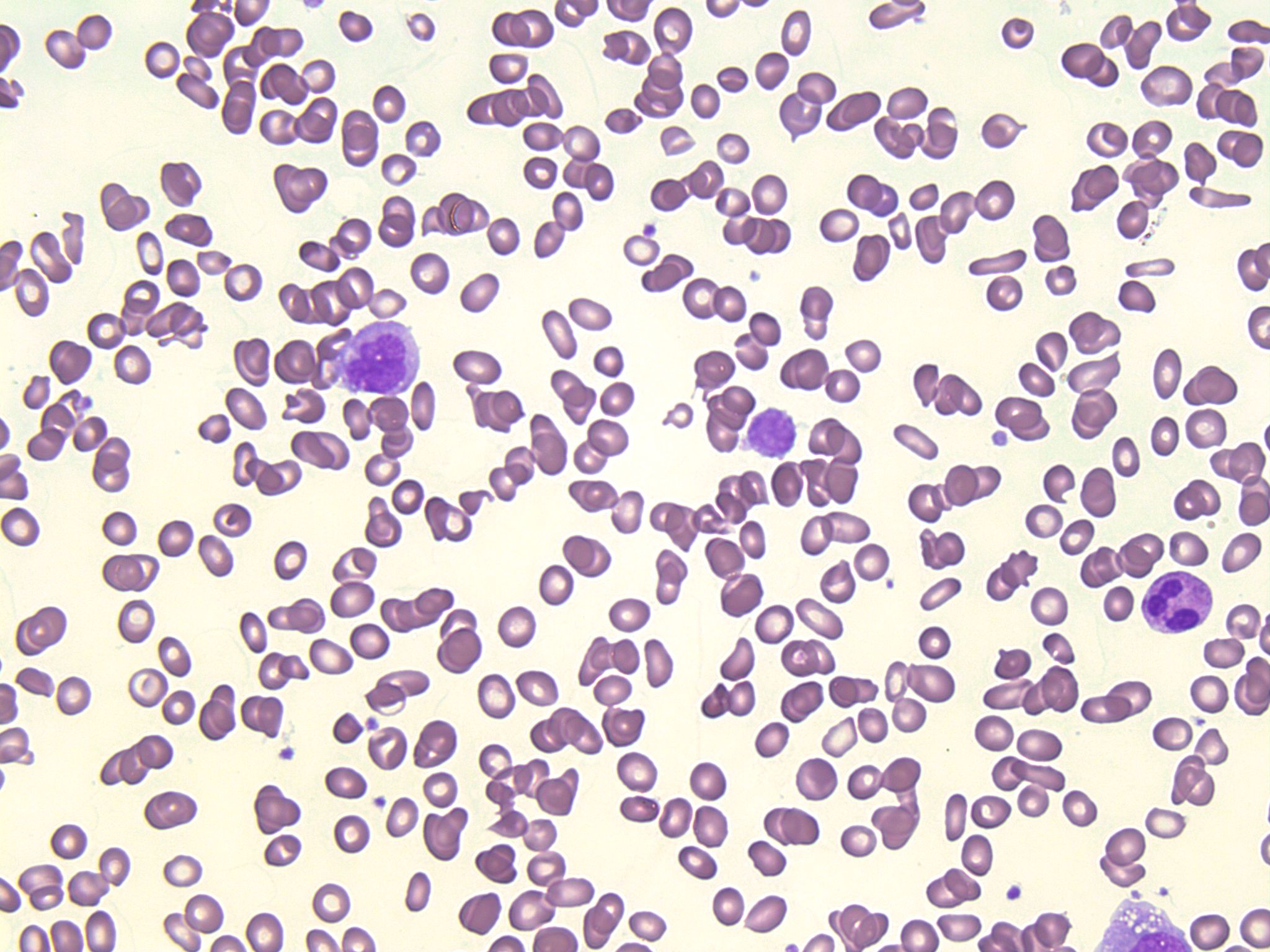
- An image from a peripheral blood smear showing tears, elliptocytes, schistocytes, and a giant platelet seen in primary myelofibrosis. 50x oil immersion. From MLS Collection, University of Alberta, https://doi.org/10.7939/R3CJ88201
-

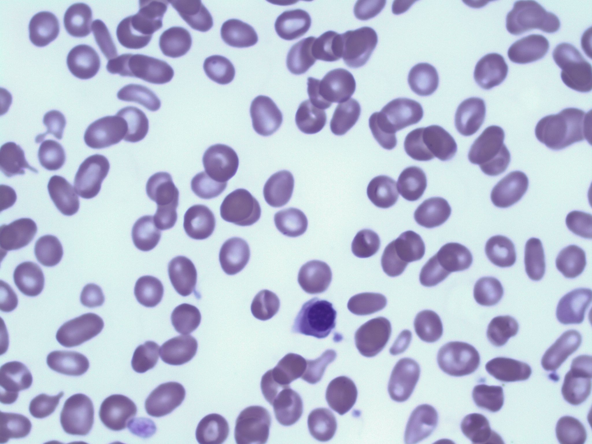
- An image from a peripheral blood smear showing tears, elliptocytes, and schistocytes, and a nucleated red blood cell seen in primary myelofibrosis. 100x oil immersion. From MLS Collection, University of Alberta, https://doi.org/10.7939/R3416TF2F
Affected Cell Line: Granulocytes and Megakaryocytes in the bone marrow resulting in secondary fibroblast stimulation and fibrotic desposition in the bone marrow.1,2
Mutation: JAK 2 V617F, CALR, and MPL gene mutations.1,3
Age Group Affected: >50 years old, occurs equally between males and females.4
Features:
Primary myelofibrosis is characterized by two stages:
1. Prefibrotic Stage
The bone marrow is hypercellular and shows minimal reticulin and fibrosis initially, with an increase in megakaryocytes and granulocytes.1
2. Fibrotic Stage
Peripheral blood shows a characteristic leukoerythroblastic picture (immature granulocyte and erythrocyte precursors) with poikilocytosis, especially teardrop cells and elliptocytes.1
Bone marrows shows marked fibrosis.1 Extramedullary hematopoiesis is often seen, with cells accumulating in the spleen, liver, and other organs.
Laboratory Findings for Primary Myelofibrosis:2,4,5
|
CBC: Early Stage: RBC: Normal WBC: Increased PLT: Increased Hb: Normal
Fibrotic Stage: RBC: Decreased WBC:Decreased PLT: Decreased Hb: Decreased |
PBS: Platelets have a dysplastic morphology (Giant, agranular) May see micromegakaryocytes Variable poikilocytosis
Fibrotic Stage: Pancytopenia Leukoerythroblastic picture Teardrop cells Elliptocytes nRBCs |
BM: Often results in a dry tap Hypercellular Fibrosis of varying degrees (Marked fibrosis in later stages) Megakaryocyte aggregates Dysgranulopoiesis Dysmegakaryopoiesis |
|
Other Tests: PLT Function: Abnormal |
References:
1. Swerdlow SH, Campo E, Harris NL, Jaffe ES, Pileri SA, Stein H, et al. editors. WHO Classification of Tumours of Haematopoietic and Lymphoid Tissues Volume 2. 4th ed. International Agency for Research on Cancer (IARC); 2008.
2. Schaub CR. Chronic Myeloproliferative disorders I: chronic myelogenous leukemia. In: Clinical hematology and fundamentals of hemostasis. 5th ed. Philadelphia: F.A. Davis Company; 2009. p. 371-84.
3. Choi CW, Bang S-M, Jang S, Jung CW, Kim H-J, Kim HY, et al. Guidelines for the management of myeloproliferative neoplasms. Korean J Intern Med [Internet]. 2015 Nov 30 [cited 2018 Jul 9];30(6):771–88. Available from: http://kjim.org/journal/view.php?doi=10.3904/kjim.2015.30.6.771
4. Randolph TR. Myeloproliferative neoplasms. In: Clinical laboratory hematology. 3rd ed. New Jersey: Pearson; 2015. p. 450-78.
5. Randolph TR. Myeloproliferative neoplasms. In: Rodak’s hematology clinical applications and principles. 5th ed. St. Louis, Missouri: Saunders; 2015. p.561-90.
