
19 Cabot Rings
Michelle To and Valentin Villatoro
-

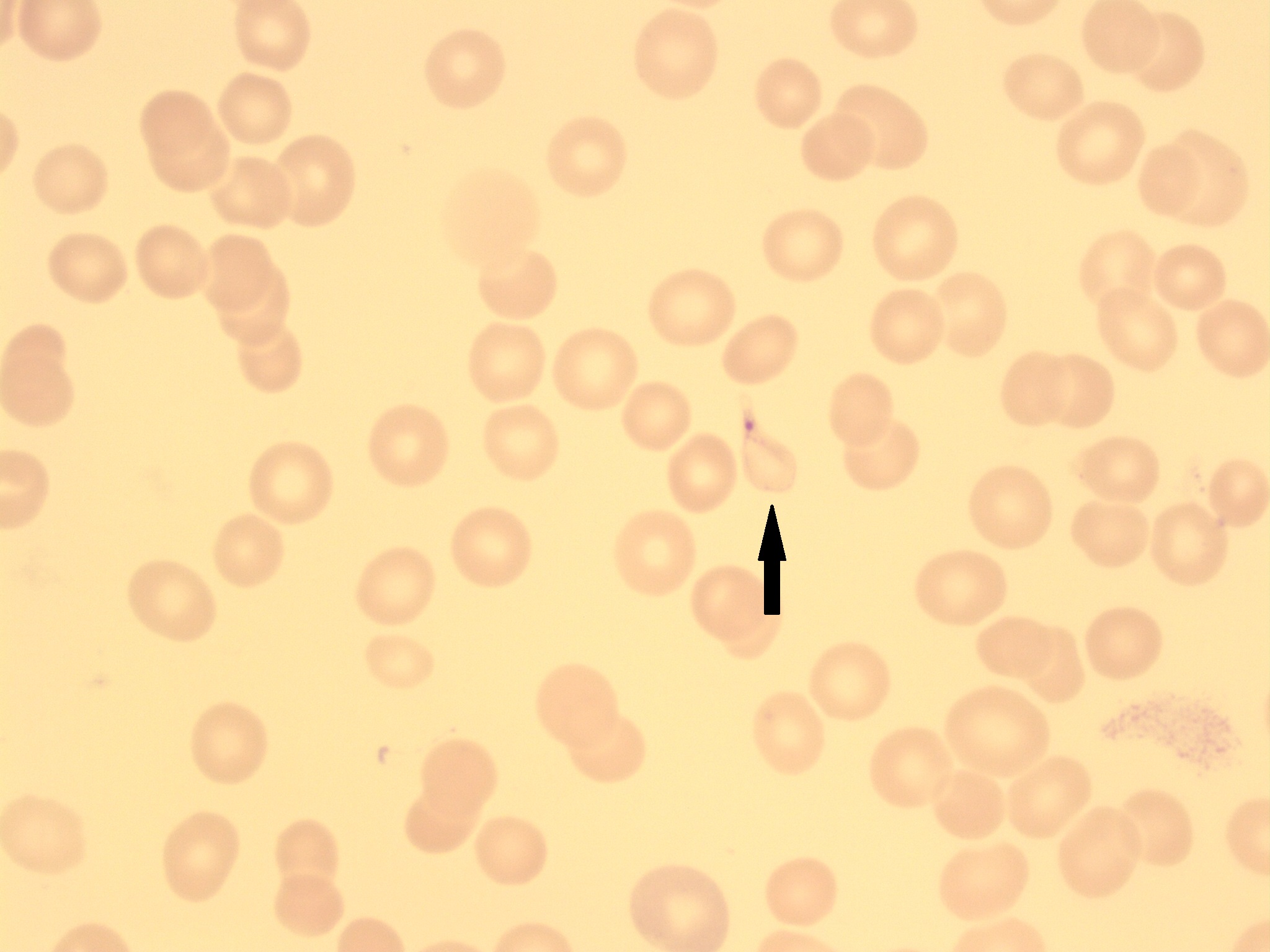
- An image from a peripheral blood smear showing a cabot ring. 100x oil immersion. From MLS Collection, University of Alberta, https://doi.org/10.7939/R3B854027
Appearance:
Red-purple inclusions that appear as a loop, ring, or figure-eight shape and span the diameter of the red blood cell. 1-2 cabot rings may be seen in a single cell.1
Note: Finding is rare, and not to be confused with malaria.
Inclusion composition:1
Remnant microtubules of mitotic spindle
Associated Disease/Clinical States:1-3
Myelodysplastic Syndrome (MDS; Dyserythropoiesis)
Megaloblastic Anemia
Lead poisoning
References:
1. Rodak BF, Carr JH. Inclusions in erythrocytes. In: Clinical hematology atlas. 5th ed. St. Louis, Missouri: Elsevier Inc.; 2017. p. 107-14.
2. Landis-Piwowar K, Landis J, Keila P. The complete blood count and peripheral blood smear evaluation. In: Clinical laboratory hematology. 3rd ed. New Jersey: Pearson; 2015. p. 154-77.
3. Turgeon ML. Erythrocyte morphology and inclusions. In: Clinical hematology: theory and procedures. 4th ed. Philadelphia, PA: Lippincott Williams & Wilkins; 1999. p. 99-111.
