
64 Toxic Changes
Michelle To and Valentin Villatoro
-
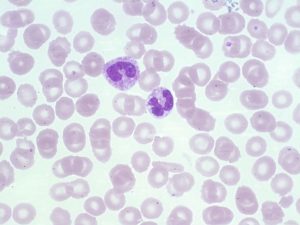
- An image from a peripheral blood smear showing a band with a blue dohle body inclusion found in the center of the cell and toxic granulation. 100x oil immersion. From MLS Collection, University of Alberta, https://doi.org/10.7939/R3930P95D

-
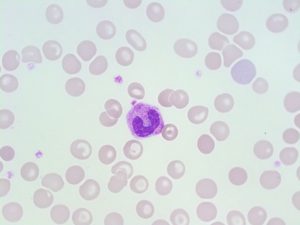
- An image from a peripheral blood smear showing toxic changes (Granulation, vacuolation) in two neutrophils. 100x oil immersion. From MLS Collection, University of Alberta, https://doi.org/10.7939/R3891261T

-
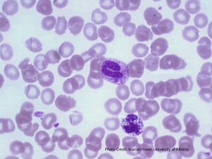
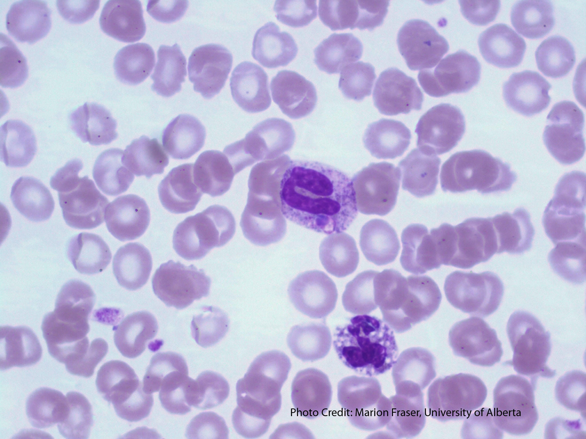
- A peripheral blood smear picture showing granulocytes with toxic changes. The Band (center) shows toxic granulation and a dohle body. The neutrophil (bottom) shows toxic vacuolation. From MLS Collection, University of Alberta, https://doi.org/10.7939/R3HT2GT1K
Cell Features:1,2
Toxic morphological changes are seen in neutrophils. A left shift with an increase in immature granulocytes typically accompanies toxic changes. In order to report toxic changes, typically two out of the three features should be seen in the majority of neutrophils:
1. Toxic Granulation:1-3
Dark blue-black peroxidase positive granules that appear in the cytoplasm of the neutrophil. Appear very similar to Alder-Reilly bodies found in Alder-Reilly anomaly but is commonly found with other features of toxicity. Can be found in mature neutrophils, bands, and metamyelocytes.
2. Toxic Vacuolation:1-3
Clear, circular, and unstained cytoplasmic areas that represent phagocytosis or autophagocytosis. Vacuoles may contain bacteria or yeast if the patient is septic.
3. Dohle Bodies:1-3
Pale blue, round or elongated cytoplasmic inclusions containing remnant ribosomal ribonucleic acid (RNA) in parallel rows (rough endoplasmic reticulum). Often present in mature neutrophils and bands near the periphery of the cell. Bodies are non-specific and can appear in several conditions such as pregnancy, cancer, burns, and infections.
Note: A left shift is usually seen on the peripheral blood smear when toxicity is present. A Left shift refers to the increase presence of immature bands and myeloid precusors.
Cause:1
Reaction to infection, inflammation, stress, and granulocyte colony-stimulating factor therapy
Laboratory Features:1,2
|
CBCD: Moderate leukocytosis Neutrophilia |
Peripheral Blood Smear: At least 2 of 3 toxic changes in the majority of neutrophils Left shift (often) |
References:
1. Marionneaux S. Nonmalignant leukocyte disorders. In: Rodak’s hematology clinical applications and principles. 5th ed. St. Louis, Missouri: Saunders; 2015. p. 475-97.
2. Jones KW. Evaluation of cell morphology and introduction to platelet and white blood cell morphology. In: Clinical hematology and fundamentals of hemostasis. 5th ed. Philadelphia: F.A. Davis Company; 2009. p. 93-116.
3. Landis-Piwowar K. Nonmalignant disorders of leukocytes: granulocytes and monocytes. In: Clinical laboratory hematology. 3rd ed. New Jersey: Pearson; 2015. p. 388-407.
