
47 Infectious Agents
Michelle To and Valentin Villatoro
Malarial Infection
-
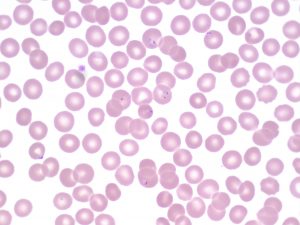
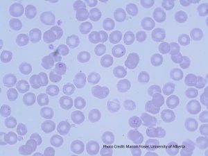
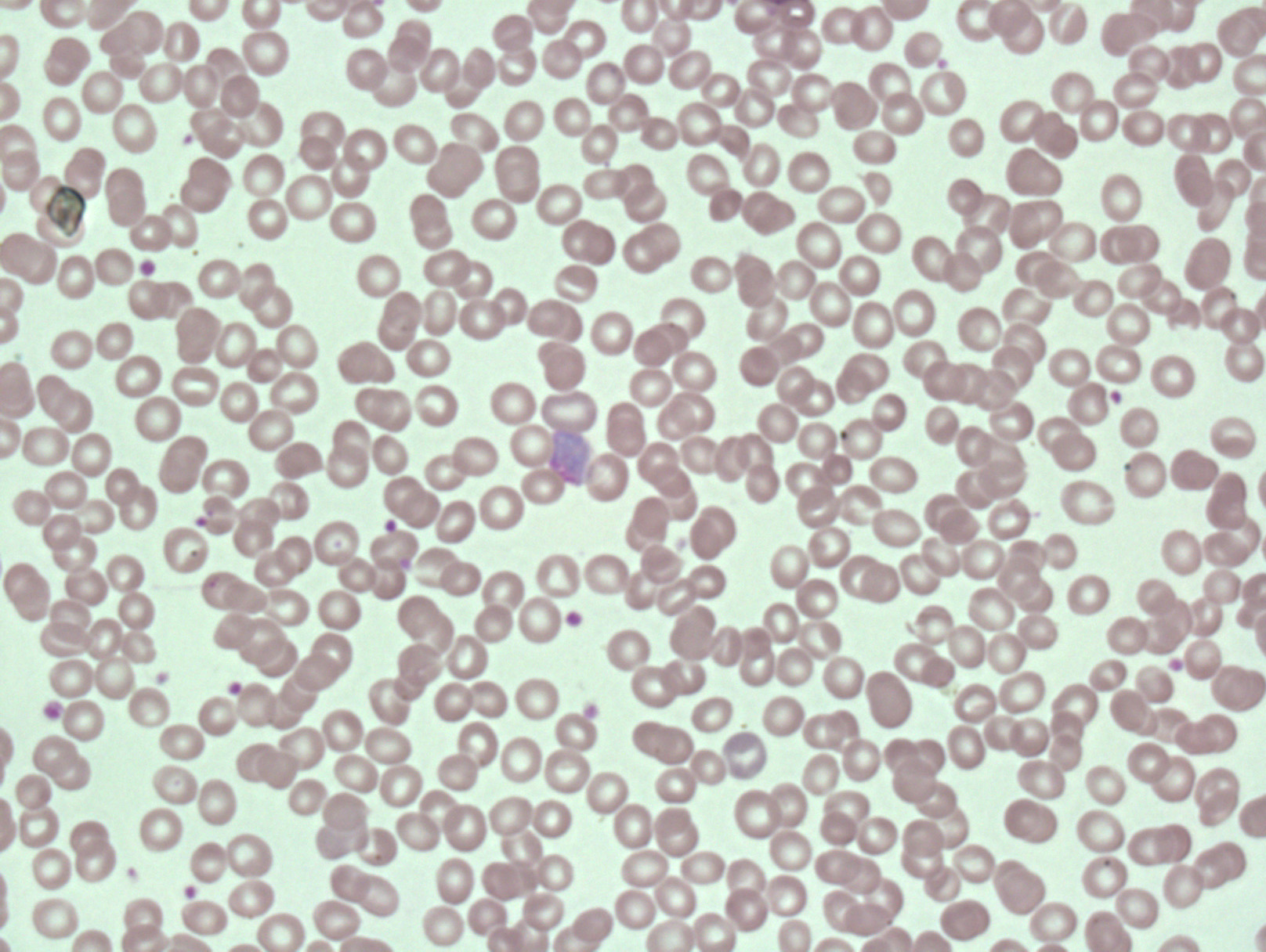
- An image from a peripheral blood smear demonstrating intra-cellular malarial rings (Plasmodium falciparum) inside red blood cells. From MLS Collection, University of Alberta, https://doi.org/10.7939/R34J0BC8J
-
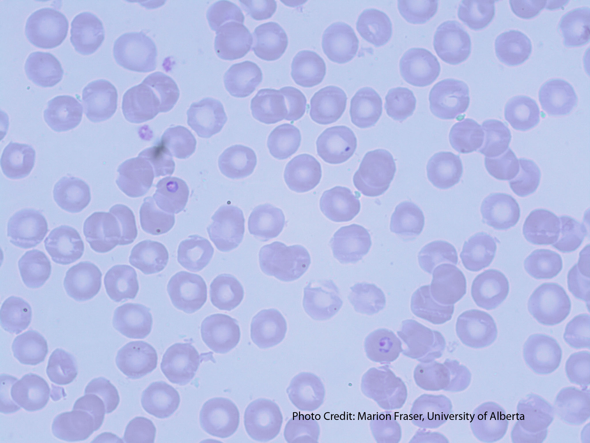
- An image from a peripheral blood smear showing two crescent-shaped gametocytes, characteristic of Plasmodium falciparum. From MLS Collection, University of Alberta, https://doi.org/10.7939/R3BG2HS02
-
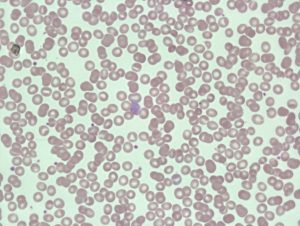
- An image from a peripheral blood smear showing ring forms of Plasmodium ovale in oval-shaped red blood cells. From MLS Collection, University of Alberta, https://doi.org/10.7939/R3W08WX95
-

- An image from a peripheral blood smear demonstrating the gametocyte of Plasmodium vivax. From MLS Collection, University of Alberta, https://doi.org/10.7939/R3R78645T
-
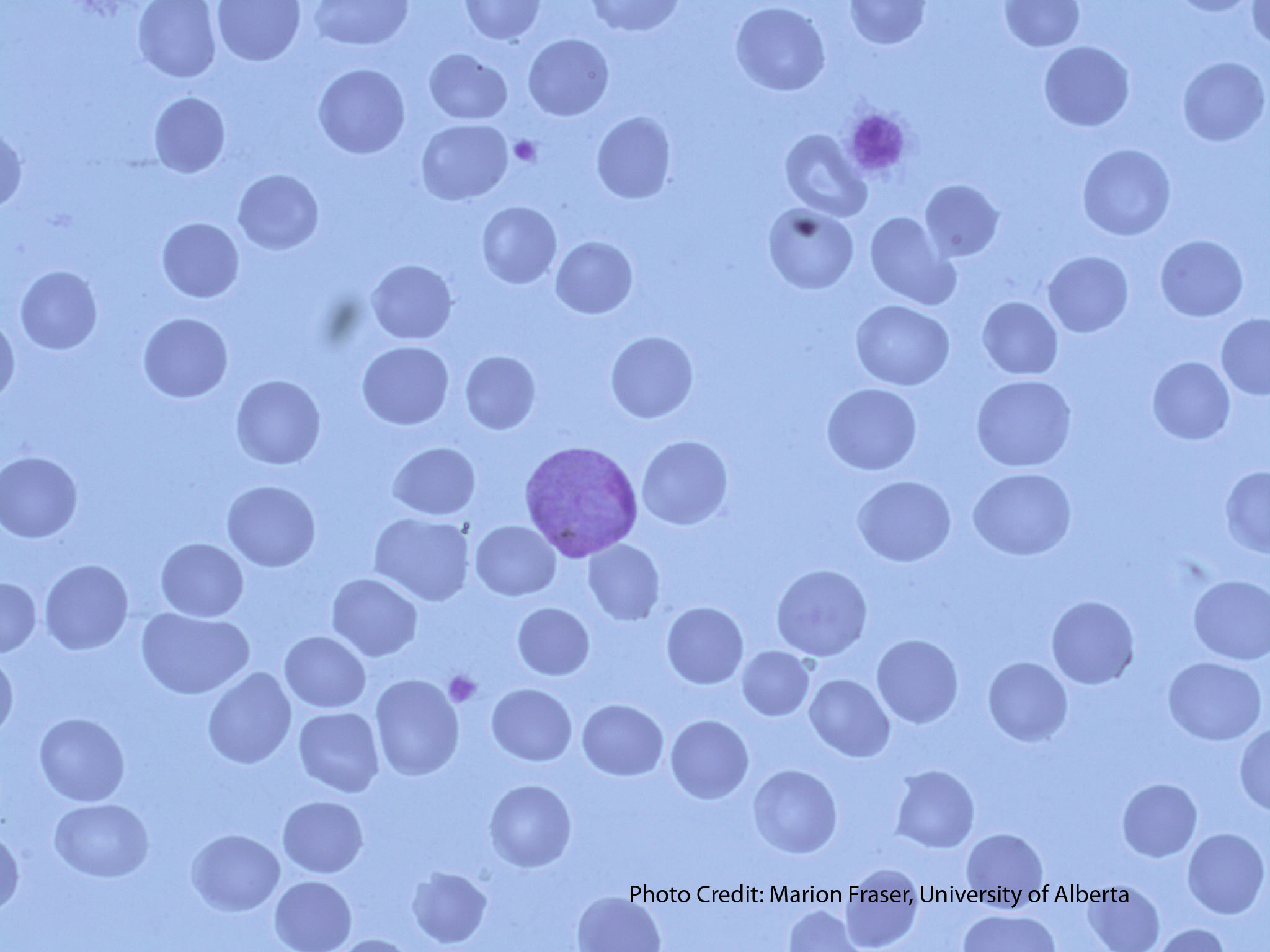
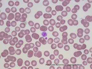
- An image from a peripheral blood smear demonstrating malaria at the gametocyte stage in the center. 60X oil immersion. From MLS Collection, University of Alberta, https://doi.org/10.7939/R33J39G6B
-
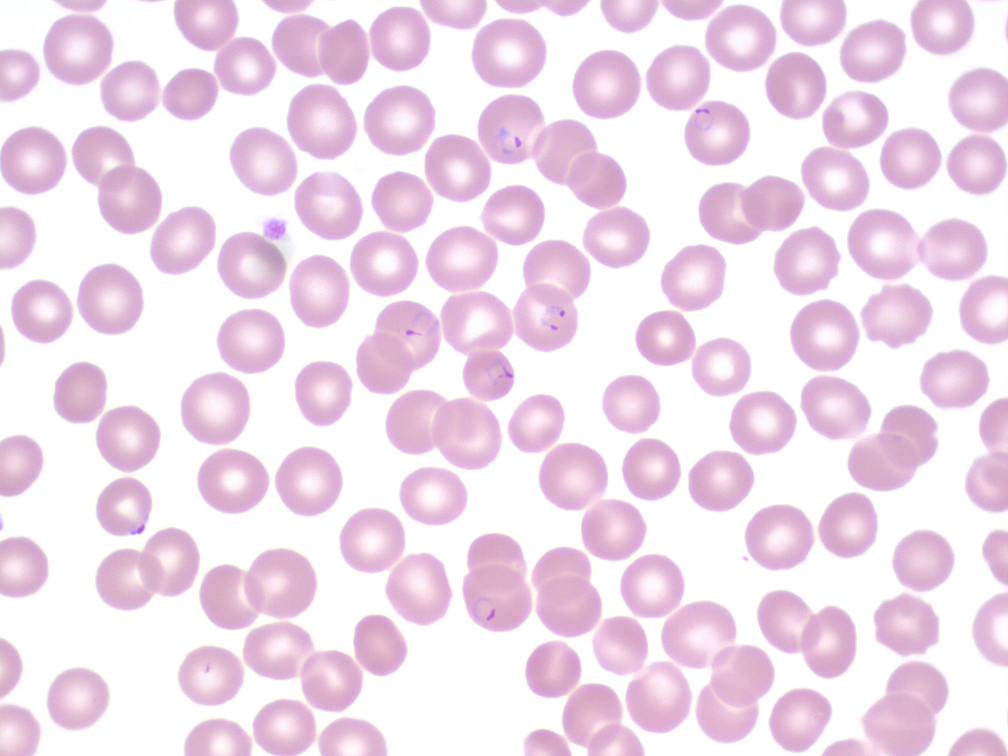
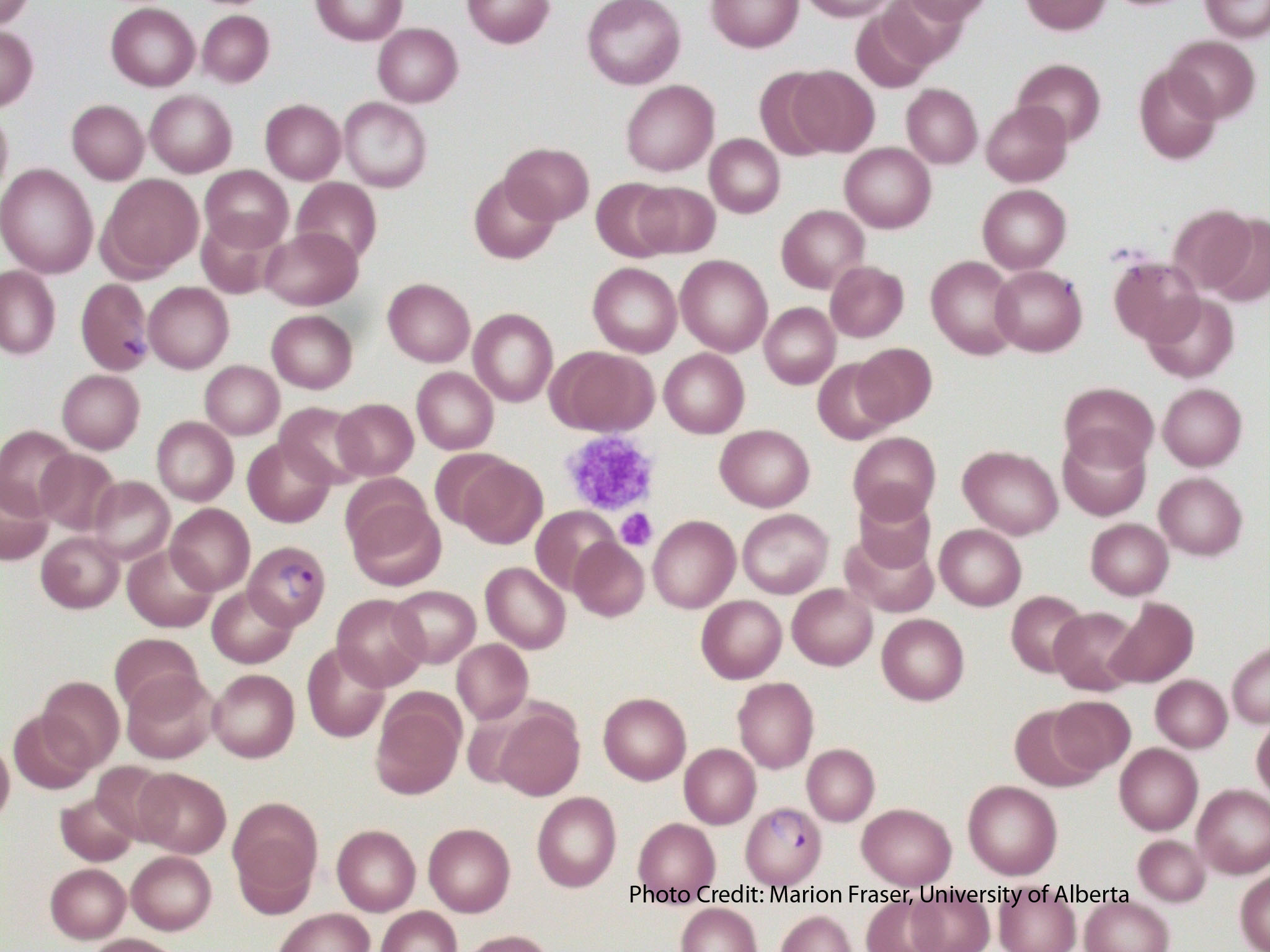
- An image from a peripheral blood smear showing signet ring forms of malaria and a large platelet in the center. From MLS Collection, University of Alberta, https://doi.org/10.7939/R3MG7GB25
Causative Agent:1,2
Malaria is transmitted by the female Anopheles mosquito.
There are five species of malaria:
Plasmodium falciparum – Most severe
Plasmodium vivax – Most common
Plasmodium ovale – Less common
Plasmodium malariae – Less common
Plasmodium knowlesi
Geographic Distribution:1
Tropical and subtropical areas
Clinical Features:1,2
Fever, headache, chills, sweating, splenomegaly. Patients can show cyclical patterns of fever and chills being present then absent. In severe malaria, jaundice, shock, bleeding, seizures, or coma may occur. Thrombocytopenia commonly occurs with malarial infections, and may lead to bleeding tendencies.
Hemolysis is mostly intravascular and occurs through a number of different mechanisms:1,2
-
Direct damage to the red blood cells
-
Immune activation of macrophage activity (Increased extravascular hemolysis)
-
Release of cytokines that inhibit erythropoiesis (inhibit erythropoietin)
-
Increased oxidative stress on RBCs during infection
-
Induction of apoptosis in red blood cell precursors
Laboratory Results for Malarial Infections:1,2
|
CBC: RBC: Decreased WBC: Normal to Slightly Increased (neutropenia during chills) PLT: Decreased Hct: Decreased RETIC: Decreased |
PBS: Normochromic Normocytic Intracellular Malarial parasite forms (forms and stages of development seen differ by species) |
BM: Erythroid Hypoplasia |
|
Other Tests: Giemsa stain Thin Smears: Morphology and parasitemia calculation Thick Smears: Presence or absence of malaria Immunoassays Flow Cytometry Molecular Testing (PCR) |
Note: See “Malaria” under RBC inclusions for additional information.
Babesiosis
Causative agents:1,3
Babesia microti is carried by the Ixodes scapularis ticks. Babesia can also be transmitted by blood transfusions.
Geographical Distribution:3
Northeast and Midwest United States
Clinical Features:3
Clinical symptoms include: flu-like symptoms ranging from fever, headache, anorexia, fatigue. Some patients may be asymptomatic. Hepatosplenomegaly may or may not be present.
Laboratory Results for Babesiosis:1,3
|
CBC: WBC: Decreased PLT: Decreased Hb: Decreased RETIC: Increased |
PBS: RBCs shows tetrads of merozoites (Maltese cross) |
Other Tests: Hemoglobinuria Proteinuria Immunoassays Molecular testing (PCR) |
Note: See “Babesia” under Abnormal RBC inclusions for additional information.
Clostridial Sepsis
Causative agent:1,3
Clostridium perfringens is an anaerobic, gram-positive spore-forming bacilli that can infect tissues after events involving trauma to the body. The bacteria releases enzymes and toxins that damage the surrounding tissue and can allow the bacteria to reach the bloodstream.
Blood infection is often fatal and can lead to massive hemolysis and the activation of DIC.
Mechanism of RBC Destruction:1,3
Hemolysis by C. perfringens is intravascular and can occur due to direct effects of alpha-toxin that it releases on the red blood cell membrane. Cell becomes spherical and subject to osmotic lysis. Significant intravascular hemolysis ensues, leading to a marked decrease in hematocrit, and dark red/brown plasma and urine.
Laboratory Results for Clostridia Infections:1,3
|
CBC: RBC: Decreased PLT: Decreased Hct: Very Decreased |
PBS: Spherocytes Microcytes Ghost cells Toxic changes with a left shift +/- ingested bacteria within neutrophils |
Other Tests: Hemoglobinemia Hemoglobinuria |
References:
1. Harmening DM, Yang D, Zeringer H. Hemolytic anemias: extracorpuscular defects. 5th ed. Philadelphia: F.A. Davis Company; 2009. p. 250-79).
2. Akinosoglou KS, Solomou EE, Gogos CA. Malaria: a haematological disease. Hematology [Internet]. 2012 Mar 1 [cited 2018 Jul 9];17(2):106–14. Available from: http://www.tandfonline.com/doi/full/10.1179/102453312X13221316477336
3. Keohane EM. Extrinsic defects leading to increased erythrocyte destruction – nonimmune causes. In: Rodak’s hematology clinical applications and principles. 5th ed. St. Louis, Missouri: Saunders; 2015. p. 394-410.
