
4.3 Musculoskeletal System Pathologies
There are a number of pathologies that can affect the musculoskeletal system, and the most common pathologies will be discussed below. Many of these pathologies can be treated with medications that will be discussed later in this chapter.
Common Pathologies
Arthritis: This common disorder involves inflammation of the joint, often resulting in significant joint pain, along with swelling, stiffness, and reduced joint mobility. There are more than 100 different forms of arthritis. Arthritis may arise from aging, damage to the articular cartilage, autoimmune diseases, bacterial or viral infections, or unknown (likely genetic) causes.
Osteoarthritis (OA): Osteoarthritis is the most common type of arthritis and is associated with aging and “wear and tear” of the cartilage. Risk factors that may lead to osteoarthritis later in life include injury to a joint, jobs that involve physical labour, sports that require running, twisting, or throwing actions, and being overweight. These factors put stress on the cartilage that covers the surfaces of bones at the synovial joints, causing the cartilage to gradually become thinner. As the cartilage layer wears down, more pressure is placed on the bones. The joint responds by increasing production of the lubricating synovial fluid, but this can lead to swelling of the joint cavity, causing pain and joint stiffness as the articular capsule is stretched. The bone tissue underlying the damaged cartilage also responds by thickening, producing irregularities and causing the articulating surface of the bone to become rough or bumpy. Moving the joint then results in pain and inflammation.
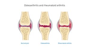
In its early stages, the symptoms of osteoarthritis may be reduced by mild activity that “warms up” the joint, but the symptoms may worsen following exercise. In individuals with more advanced osteoarthritis, the affected joints can become more painful and therefore difficult to use effectively, resulting in decreased mobility. There is no cure for osteoarthritis, but several treatments can help alleviate the pain. These treatments may include lifestyle changes such as weight loss and low-impact exercise and over-the-counter or prescription medications that help alleviate the pain and inflammation. For severe cases, arthroplasty may be required. See Fig. 4.6 for a comparison of a normal joint, a joint with osteoarthritis, and a joint with rheumatoid arthritis.
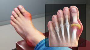
Gout: This form of arthritis results from the deposition of uric acid crystals in a joint (Fig. 4.7). Usually only one or a few joints are affected, such as the big toe, knee, or ankle. The attack may only last a few days, but may return to the same or another joint. Gout occurs when the body makes too much uric acid or the kidneys do not properly excrete it. A diet with excessive fructose has been associated with an increased chance of developing gout.
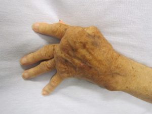
Rheumatoid arthritis (RA): This type of arthritis is an autoimmune disease that results in the joint capsule and synovial membrane becoming inflamed (Fig. 4.8). As the disease progresses, the articular cartilage is severely damaged or destroyed, resulting in joint deformation, loss of movement, and severe disability. The most commonly involved joints are the hands, feet, and cervical spine, with corresponding joints on both sides of the body usually affected. Rheumatoid arthritis is also associated with lung fibrosis, vasculitis, coronary heart disease, and premature mortality. With no known cure, treatments are aimed at alleviating symptoms.
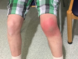
Bursitis: This condition is the inflammation of a bursa near a joint (Fig. 4.9), which causes pain, swelling, or tenderness of the bursa and surrounding area, and may also result in joint stiffness. Bursitis is most commonly associated with the bursae found at or near the shoulder, hip, knee, or elbow joints. It can be either acute (lasting only a few days) or chronic and can arise from muscle overuse, trauma, excessive or prolonged pressure on the skin, rheumatoid arthritis, gout, or infection of the joint. Repeated acute episodes of bursitis can result in a chronic condition.
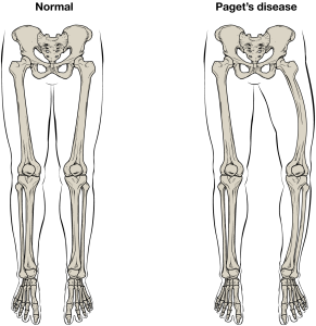
Paget’s disease: This condition usually occurs in adults over the age of 40. It is a disorder of the bone remodelling process that begins with overactive osteoclasts. This essentially means more bone is resorbed than is laid down. The osteoblasts try to compensate, but the new bone they lay down is weak and brittle and therefore prone to fracture. Although some people with Paget’s disease have no symptoms, others experience pain, bone fractures, and bone deformities as shown in Fig. 4.10. Bones of the pelvis, skull, spine, and legs are the most commonly affected. When Paget’s disease occurs in the skull, it can cause headaches and hearing loss. It is not known what makes the osteoclasts become overactive. Hereditary may play a role, and some scientists believe Paget’s disease is caused by an as-yet-unidentified virus.
Osteoporosis (OP): This disease is characterized by a decrease in bone mass that occurs when the rate of bone resorption exceeds the rate of bone formation, a common occurrence as the body ages (Fig. 4.11). Note how this is different from Paget’s disease. In Paget’s disease, new bone is formed in an attempt to keep up with the resorption by overactive osteoclasts, but the new bone is produced haphazardly. In fact, when a physician is evaluating a patient with thinning bones, they will test for osteoporosis and Paget’s disease, as well as other diseases. Osteoporosis does not have the elevated blood levels of alkaline phosphatase found in Paget’s disease.
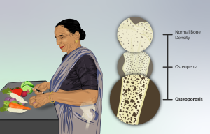
Although osteoporosis can involve any bone, it most commonly affects the proximal ends of the femur, vertebrae, and wrists. As a result of the loss of bone density, the osseous tissue may not provide adequate support for everyday functions, and something as simple as a sneeze can cause a vertebral fracture. When an elderly person falls and breaks a hip, it is very likely the femur that breaks first, which results in the fall. Histologically, osteoporosis is characterized by a reduction in the thickness of compact bone and an increase in the number and size of trabeculae in cancellous (spongy) bone.
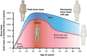
Women lose bone mass more quickly than men starting at about 50 years of age (Fig. 4.12). This occurs because 50 is the approximate age at which women go through menopause. Not only do their menstrual periods lessen and eventually cease, but their ovaries reduce in size and then cease to produce estrogen, a hormone that promotes osteoblast activity and the production of bone matrix. Thus, osteoporosis is more common in women than in men, but men can develop it, too. Anyone with a family history of osteoporosis has a greater risk of developing the disease, so the best treatment is prevention, which should start with a childhood diet that includes adequate intake of calcium and vitamin D and a lifestyle that includes weight-bearing exercise. These actions, as discussed above, are important for building bone mass. Promoting proper nutrition and weight-bearing exercise early in life can maximize bone mass before the age of 30, thus reducing the risk of osteoporosis at a later age.
For many elderly people, a hip fracture can be life threatening. The fracture itself may not be serious, but the reduced mobility required by the healing process can lead to the formation of blood clots that may lodge in the capillaries of the lungs, resulting in respiratory failure, pneumonia caused by the lack of poor air exchange that accompanies immobility, pressure sores (bed sores) that allow pathogens to enter the body and cause infections, and urinary tract infections from catheterization.
Current treatments for managing osteoporosis include medications, which will be discussed further in the next section, and minimizing the risk of falls by removing tripping hazards, for example.
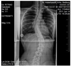
Scoliosis: This pathology is an abnormal, lateral curvature of the vertebral column, accompanied by twisting. Fig.4. 13 shows an X-ray of a young girl diagnosed with scoliosis. Compensatory curves may also develop in other areas of the vertebral column to help maintain the position of the head over the feet. Scoliosis is the most common vertebral abnormality among girls. The cause is usually unknown, but it may result from weakness of the back muscles, defects such as differential growth rates in the right and left sides of the vertebral column, or differences in the length of the lower limbs. When present, scoliosis tends to get worse during adolescent growth spurts. Although most individuals do not require treatment, a back brace may be recommended for growing children. In extreme cases, surgery may be required.
Tendinitis: This condition is characterized by inflammation of a tendon, which is the thick band of fibrous connective tissue that attaches a muscle to a bone, and causes pain and tenderness in the area around a joint. On rare occasions, a sudden serious injury will cause tendinitis. Most often, the condition results from repetitive motions over time that strain the tendons needed to perform the tasks. People whose jobs, sports, or hobbies involve performing the same movements over and over again are often at the greatest risk of tendinitis—you may have heard of tennis and golfer’s elbow, jumper’s knee, and swimmer’s shoulder. In all cases, overuse of the joint causes a microtrauma that initiates the inflammatory response. Tendinitis is routinely diagnosed through a clinical examination. In cases of severe pain, X-rays can be examined to rule out the possibility of a bone injury. Severe cases of tendinitis can even tear loose a tendon. Surgical repair of a tendon is painful, and because connective tissue in the tendon does not have an abundant blood supply, the healing process is slow. Treatment may involve physical therapy, medications, and preventive measures to decrease the chance of reoccurrence.
Now that we have reviewed the musculoskeletal system and its common pathologies, we will now discuss medication-related treatment options for these conditions.
Attribution
Unless otherwise indicated, material on this page has been adapted from the following resource:
Ernstmeyer, K., & Christman, E. (Eds.). (2020). Nursing pharmacology. Chippewa Valley Technical College. https://wtcs.pressbooks.pub/pharmacology/, licensed under CC BY 4.0
Image Credits (images are listed in order of appearance)
Osteoarthritis and rheumatoid arthritis – Normal joint Osteoarthr – Smart-Servier by Laboratoires Servier, CC BY-SA 3.0
Gout Signs and Symptoms by Scientific Animations, CC BY-SA 4.0
Rheumatoid Arthritis by James Heilman, MD, CC BY-SA 3.0
Akute Bursitis präpatellaris by Olaf Schmale, CC BY-SA 4.0
610 Feature Pagets Disease by OpenStax College, CC BY 3.0
Depiction of an Osteoporosis patient by myUpchar, CC BY-SA 4.0
615 Age and Bone Mass by OpenStax College, CC BY 3.0
Scoliosis (15-year-old) by JanDerChemiker, CC BY-SA 3.0
joint replacement surgery
inflammation of the blood vessels
cells that eat away bone
bone-building cells

.jpg)