
6.2 Cardiovascular System
The cardiovascular system uses blood to deliver nutrients and remove wastes from the trillions of cells in the human body. The heart, which is the primary organ in this system, pumps blood throughout the body via a network of blood vessels. These three components—blood, blood vessels, and the heart—make up this complex system. Most of this section will discuss the heart as it is the most complex and integral part of this body system.
The Heart
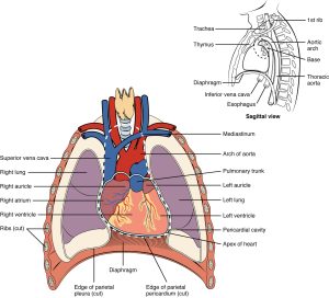
The human heart is located within the thoracic cavity, medially between the lungs in the space known as the mediastinum. The great veins, the superior vena cava and the inferior vena cava, and the great arteries, the aorta and the pulmonary trunk, are attached to the superior surface of the heart, called the base. The base of the heart is located at the level of the third costal cartilage, as seen in Figure 6.1. The inferior tip of the heart, the apex, lies just to the left of the sternum between the junction of the fourth and fifth ribs.
Chambers and Circulation Through the Heart
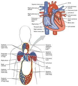
The heart consists of four chambers: two atria and two ventricles. The right atrium receives deoxygenated blood from the systemic circulation, and the left atrium receives oxygenated blood from the lungs. The atria contract to push blood into the lower chambers, the right ventricle and the left ventricle. The right ventricle contracts to push blood into the lungs, and the left ventricle is the primary pump that propels blood to the rest of the body.
There are two distinct but linked circuits in the human circulation called the pulmonary and systemic circuits. The pulmonary circuit transports blood to and from the lungs, where it picks up oxygen and delivers carbon dioxide for exhalation. The systemic circuit transports oxygenated blood to virtually all the tissues of the body and returns deoxygenated blood and carbon dioxide to the heart to be sent back to the pulmonary circulation. Figure 6.2 is an illustration of blood flow through the heart and blood circulation throughout the body.
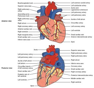
Blood also circulates through the coronary arteries with each beat of the heart. The left coronary artery distributes blood to the left side of the heart, and the right coronary artery distributes blood to the right atrium, portions of both ventricles, and the heart conduction system. See Figure 6.3 for an illustration of the coronary arteries. When a patient has a myocardial infarction, a blood clot lodges in one of the coronary arteries that perfuse the heart tissue. If a significant area of muscle tissue dies from lack of perfusion, the heart is no longer able to pump.
Conduction System of the Heart
Contractions of the heart are stimulated by the electrical conduction system. The components of the cardiac conduction system include the sinoatrial (SA) node, the atrioventricular (AV) node, the left and right bundle branches, and the Purkinje fibres. Figure 6.4 is an image of the conduction system of the heart.
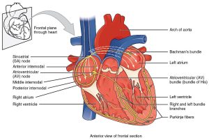
Normal cardiac rhythm is established by the sinoatrial (SA) node. The SA node has the highest rate of depolarization and is known as the pacemaker of the heart. It initiates the sinus rhythm, the normal electrical pattern followed by heart contractions. The SA node initiates the action potential, which sweeps across the atria through the AV node to the bundle branches and Purkinje fibres, and then spreads to the contractile fibres of the ventricle to stimulate contraction of the ventricle (Betts et al., 2013).
Cardiac Cycle and Output
The period of time that begins with contraction of the atria and ends with ventricular relaxation is known as the cardiac cycle. The period of contraction that the heart undergoes while it pumps blood into circulation is called systole. The period of relaxation that occurs as the chambers fill with blood is called diastole. A common and important unit of measurement is cardiac output, which is a measurement of the amount of blood pumped by each ventricle in one minute.
Heart Rate
Heart rate (HR) can vary considerably, not only with exercise and fitness levels, but also with age. A newborn’s resting heart rate may be 120 to 160 beats per minute (bpm). Heart rate gradually decreases until young adulthood, then gradually increases with age. For an adult, a normal resting HR will be in the range of 60 to 100 bpm. Bradycardia is the condition in which the resting heart rate drops below 60 bpm, and tachycardia is the condition in which the resting heart rate is above 100 bpm.
Blood Vessels
After blood is pumped out of the ventricles, it is carried through the body via blood vessels. An artery is a blood vessel that carries blood away from the heart. From there, the artery branches into ever-smaller vessels and eventually into tiny capillaries, where nutrients and wastes are exchanged at the cellular level. Capillaries then combine with other small blood vessels that carry blood to a vein, a larger blood vessel that returns blood to the heart. Compared to arteries, veins are thin-walled, low-pressure vessels. Larger veins are also equipped with valves that promote the one-way flow of blood towards the heart and prevent backflow caused by the inherent low blood pressure in veins as well as the pull of gravity.
In addition to their primary function of returning blood to the heart, veins may be considered blood reservoirs because systemic veins contain approximately 64% of the blood volume at any given time. Approximately 21% of the venous blood is located in venous networks within the liver, bone marrow, and integument. This volume of blood is referred to as the venous reserve. Through venoconstriction, this reserve volume of blood can be returned to the heart quickly for redistribution to other parts of the cardiovascular system.
The following video provides an overview of the cardiovascular system and summarizes much of the content covered in this section.
(Alila Medical Media, 2019)
Attribution
Unless otherwise indicated, material on this page has been adapted from the following resource:
Betts, J. G., Young, K. A., Wise, J. A., Johnson, E., Poe, B., Kruse, D. H., Korol, O., Johnson, J. E., Womble, M., & DeSaix, P. (2013). Anatomy and physiology. OpenStax. https://openstax.org/details/books/anatomy-and-physiology, licensed under CC BY 4.0
References
Image Credits (images are listed in order of appearance)
2001 Heart Position in ThoraxN by OpenStax College, CC BY 3.0
2003 Dual System of Human Circulation by OpenStax College, CC BY 3.0
2005 Surface Anatomy of the Heart by OpenStax College, CC BY 3.0
2018 Conduction System of Heart by OpenStax College, CC BY 3.0
heart attack
normal sinus rhythm is a regular heartbeat
slow heart rate
increased heart rate
