
6.3 Cardiovascular System Pathologies
A number of pathologies can affect the cardiovascular system, and the more common ones will be discussed below. Many of these pathologies can initially be treated with lifestyle changes but often will require medications that will be discussed later in this chapter.
Common Pathologies
Hypertension: Chronically elevated blood pressure (an increase in both systolic and diastolic pressure) is known clinically as hypertension. High blood pressure is first treated with lifestyle changes such as diet and exercise, and if this does not work, then medication is added to the treatment plan. Approximately 6 million Canadians have hypertension (Heart and Stroke Foundation of Canada, 2015). The current guidelines state that hypertension should be treated at 130/80 mm Hg rather than the previous standard of 140/90 mm Hg.
Unfortunately, hypertension is often a “silent disorder,” meaning that no symptoms occur until complications happen, so patients may fail to recognize the seriousness of their condition and fail to follow their treatment plan. The result is often a heart attack or stroke. Hypertension may also lead to an aneurysm, peripheral artery disease, chronic kidney disease, or heart failure (Betts et al., 2013).
Medications commonly used to treat hypertension include diuretics, ACE inhibitors, beta-blockers, and calcium channel blockers. These medications will be discussed in detail in the next section.
The following video provides an overview on the topic of hypertension.
(Alila Medical Media, 2018)
Hyperlipidemia: Cholesterol is a fat that your body needs to work properly. However, too much “bad” cholesterol can increase the risk for heart disease, stroke, and peripheral vascular disease. The medical term for high blood cholesterol is hyperlipidemia. A list and description of the types of cholesterol can be found in the Key Concept box below.
Key Concept
- Total cholesterol: All the cholesterols combined.
- High-density lipoprotein (HDL) cholesterol: Often called “good” cholesterol because it promotes the excretion of cholesterol. Exercise helps to increase HDL and remove cholesterol from the bloodstream.
- Low-density lipoprotein (LDL) cholesterol: Often called “bad” cholesterol because it stores cholesterol in the bloodstream, which contributes to atherosclerosis.
- Very-low-density lipoprotein (VLDL) cholesterol: Another “bad” cholesterol because it carries triglycerides that are associated with plaque formation in the arteries.
(Cleveland Clinic, 2022)
For many people, abnormal cholesterol levels are partly caused by lifestyle choices, including a diet that is high in fat, being overweight, or lack of exercise. However, disorders that lead to abnormal cholesterol and triglyceride levels can also be passed down through families (Cleveland Clinic, 2022). In addition to lifestyle modifications such as eating a low-fat diet and getting more exercise, hyperlipidemia is treated with antilipidemic medications, which will be discussed in the next section.
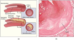
Atherosclerosis: This condition begins with injury to the endothelium of an artery, which may be caused by irritation from high blood glucose, infection, tobacco use, excessive blood lipids, and other factors. Injury to the artery walls results in inflammation, and as the inflammation spreads further into the artery wall, it weakens and scars it, making it stiff. Circulating triglycerides and cholesterol can seep between the damaged lining cells and become trapped within the artery wall, where they are joined by leukocytes, calcium, and cellular debris. Eventually, this buildup, called plaque, can narrow arteries enough to impair blood flow. The term for this condition, atherosclerosis, describes the plaque deposits. See Figure 6.5 for an illustration of atherosclerosis (Betts et al., 2013).
Sometimes a plaque can rupture, causing microscopic tears in the artery wall that allow blood to leak into the tissue on the other side. When this happens, platelets rush to the site to clot the blood. This clot can further obstruct the artery, and if this occurs in a coronary or cerebral artery, it can cause a sudden heart attack or stroke. Alternatively, plaque can also break off and travel through the bloodstream as an embolus until it blocks a more distant, smaller artery.
Even without total blockage, narrowed vessels lead to ischemia. Ischemia can lead to hypoxia, causing a myocardial infarction or cerebrovascular accident.
Treatment of atherosclerosis includes lifestyle changes, such as weight loss, smoking cessation, regular exercise, and adoption of a diet low in sodium and saturated fats. Antilipidemic medications are prescribed to reduce cholesterol and help prevent atherosclerosis and will be discussed in more detail in the next section.
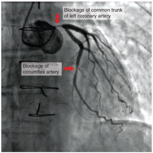
Coronary artery disease: This condition is the leading cause of death worldwide. It occurs when atherosclerosis within the walls of the coronary arteries obstructs blood flow. As the coronary blood vessels become blocked with plaque, the flow of blood to the tissues is restricted, causing the cardiac cells to receive insufficient amounts of oxygen, which can cause pain called angina. Figure 6.6 shows the blockage of coronary arteries highlighted by the injection of dye. Some individuals with coronary artery disease report pain (angina) radiating from the chest, but others, especially women, may remain asymptomatic or have alternative symptoms of neck, jaw, shoulder, upper back, or abdominal pain. If untreated, coronary artery disease can lead to a myocardial infarction, or MI. Risk factors include smoking, family history of coronary artery disease, hypertension, obesity, diabetes, lack of exercise, stress, and hyperlipidemia. Treatment may include medication, changes to diet, exercise, a coronary angioplasty with a balloon catheter, insertion of a stent, or a coronary bypass procedure (Betts et al., 2013).
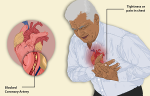
Myocardial infarction (MI): This pathology is commonly referred to as a heart attack and results from lack of blood flow and oxygen to a region of the heart, causing the death of cardiac muscle cells. An MI often occurs when a coronary artery is blocked by the buildup of atherosclerotic plaque and becomes a thrombus or when a portion of an unstable atherosclerotic plaque travels through the coronary arterial system and lodges in one of the smaller vessels.
In the case of an acute MI, there is often sudden pain, called angina, beneath the sternum (retrosternal pain), which often radiates down the left arm in male patients (see Fig. 6.7), but does not commonly do so in female patients. In addition, patients typically present with difficulty breathing and dyspnea, irregular heartbeat (palpitations), nausea and vomiting, diaphoresis, anxiety, and syncope, though not all of these symptoms may be present. Many of the symptoms are shared with other medical conditions, including anxiety attacks and simple indigestion, so accurate diagnosis is critical for patient survival.
An MI can be confirmed by examining the patient’s ECG, which frequently reveals alterations in the ST and Q components. Immediate treatment for an MI is required and includes administering supplemental oxygen, aspirin, and nitroglycerin, which will be discussed further in the next section. Longer-term treatments may include injections of thrombolytic agents, such as tPA, that dissolve the clot, along with the anticoagulant heparin, a balloon angioplasty with stents to open blocked vessels, or bypass surgery to allow blood to pass around the site of the blockage (Betts et al., 2013).
The following video is an overview of coronary artery disease and what happens during a myocardial infarction.
(FreeMedEducation, 2021)
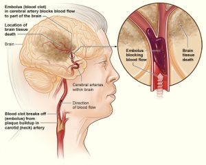
Cerebrovascular accident (CVA): A CVA results when there is lack of blood flow to part or all of the brain. The internal carotid arteries, along with the vertebral arteries, are the two primary suppliers of blood to the human brain. Given the central role and vital importance of the brain to life, it is critical that blood supply to this organ remains uninterrupted. However, blood flow may become obstructed because of atherosclerosis or an embolus that has travelled from elsewhere in the bloodstream. For example, an arrhythmia called atrial fibrillation can cause clots to form in the heart that then move to the brain. When blood flow is interrupted, even for just a few seconds, a transient ischemic attack (TIA) occurs, resulting in loss of consciousness or temporary loss of neurological function. Loss of blood flow for longer periods produces irreversible brain damage or a stroke, also called a cerebrovascular accident (Betts et al., 2013).
There are two types of cerebrovascular accidents: ischemic and hemorrhagic. Ischemic strokes are caused by atherosclerosis or a blood clot that blocks the flow of blood to the brain (see Figure 6.8). Eighty percent of strokes are ischemic. Hemorrhagic strokes are caused by a blood vessel that ruptures and bleeds into the brain. Risk factors for a stroke include smoking, high blood pressure, and cardiac arrhythmias. Treatment of a stroke depends on the cause (Betts et al., 2013). Ischemic strokes are treated with thrombolytic medication such as tPA to dissolve the clot, whereas hemorrhagic strokes often require surgery to stop the bleeding.
Key Concept
The mnemonic FAST helps people remember what to look for when someone is dealing with a sudden loss of neurological function (Betts et al., 2013). If someone complains of feeling “funny,” check these things quickly:
- Look at the person’s face. Do they have problems moving Face muscles and making regular facial expressions?
- Ask the person to raise their Arms above their head. Can the person lift one arm but not the other?
- Has the person’s Speech changed? Are they slurring words or having trouble saying things?
- If any of these things have happened, then it is Time to call for help.
(Betts et al., 2013)
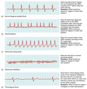
Arrhythmias: Occasionally, an area of the heart other than the SA node will initiate an impulse that is followed by a premature contraction; such an area is known as an ectopic focus. An ectopic focus may be stimulated by localized ischemia, exposure to certain drugs, elevated stimulation by both sympathetic or parasympathetic divisions of the autonomic nervous system, or one of several diseases or pathological conditions. Occasional occurrences are generally temporary and are not life-threatening, but if the condition becomes chronic, it may lead to either an arrhythmia or fibrillation. Severe arrhythmias can lead to a cardiac arrest, which is fatal if not treated within a few minutes. Abnormalities that may be detected by ECGs are shown in Figure 6.9.
Heart failure or congestive heart failure (CHF): Heart failure is a condition in which the heart can’t pump enough blood to meet the body’s needs. Right-sided heart failure occurs when the heart can’t pump enough blood to the lungs to pick up oxygen, whereas left-side heart failure occurs when the heart can’t pump enough oxygen-rich blood to the rest of the body. Heart failure is a very common condition, but there is no cure. The symptoms can be managed for several years with lifestyle modifications and different types of medications. Causes of heart failure include hypertension, myocardial infarction, and other cardiac and respiratory diseases. Common symptoms of heart failure include peripheral edema and shortness of breath that occur as a result of fluid overload. Many patients are treated with diuretics to manage the symptoms of fluid overload, anti-hypertensive medications to manage blood pressure, and medications to increase the contractility of the heart, which will be discussed in the next section.
Attribution
Unless otherwise indicated, material on this page has been adapted from the following resource:
Ernstmeyer, K., & Christman, E. (Eds.). (2020). Nursing pharmacology. Chippewa Valley Technical College. https://wtcs.pressbooks.pub/pharmacology/, licensed under CC BY 4.0
References
Alila Medical Media. (2018, February 13). Hypertension – High blood pressure animation [Video]. YouTube. https://www.youtube.com/watch?v=JtBtk00EiVM
Betts, J. G., Young, K. A., Wise, J. A., Johnson, E., Poe, B., Kruse, D. H., Korol, O., Johnson, J. E., Womble, M., & DeSaix, P. (2013). Anatomy and physiology. OpenStax. https://openstax.org/details/books/anatomy-and-physiology, licensed under CC BY 4.0
Cleveland Clinic. (2022). Cholesterol numbers and what they mean. https://my.clevelandclinic.org/health/articles/11920-cholesterol-numbers-what-do-they-mean
FreeMedEducation. (2021, January 12). What happens during a heart attack? – What is coronary heart disease? [Video]. YouTube. https://youtu.be/xBAvxnT0ZvI
Heart & Stroke Foundation of Canada. (2015). Get your blood pressure under control. https://www.heartandstroke.ca/-/media/pdf-files/canada/health-information-catalogue/en-get-your-blood-pressure-under-control-v21-web.ashx?la=en&hash=20493566724F3F87387717A720D49F60521C1CF2
Image Credits (images are listed in order of appearance)
2113ab Atherosclerosis by OpenStax College, CC BY 3.0
2016 Occluded Coronay Arteries by OpenStax College, CC BY 3.0
A man having a Heart Attack by myUpchar, CC BY-SA 4.0
Stroke ischemic by National Heart Lung and Blood Institute (NIH), Public domain
2024 Cardiac Arrhythmias by OpenStax College, CC BY 3.0
myocardial infarction; abbreviated as MI
a cerebrovascular accident (CVA)
ballooning of a blood vessel caused by weakening of the vessel wall
obstruction of blood vessels in peripheral regions of the body
the kidneys are damaged and no longer work properly
failure of the heart to pump well; the heart becomes weakened and can't supply the cells with enough blood
also known as a lipid
high lipid levels in the blood
small piece of a blood clot that breaks off; plural form is emboli
reduced blood flow to the tissue region “downstream” of a narrowed vessel
decreased supply of oxygen to the tissues
heart attack
a stroke; often abbreviated as CVA
hardened with plaque
chest pain caused by lack of blood flow (and oxygen) to cardiac cells
high blood pressure
a blood clot
shortness of breath or difficulty breathing
sweating
fainting
electrocardiogram
small pieces of a blood clot that break off; singular form is embolus
irregular heart rhythm
also know as a mini-stroke; often abbreviated as TIA
bleeding caused by blood vessel rupture
sinoatrial node, which is responsible for the contraction of the atria
an uncoordinated beating of the heart
heart stops beating suddenly
swelling of the limbs, usually the legs
