
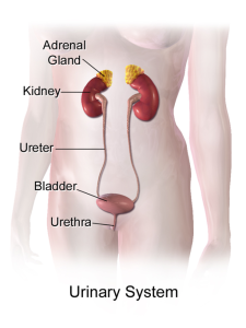
2.2 The Urinary System
The urinary system, shown in Fig. 2.1, is responsible for cleansing the blood and removing wastes from the body. However, it has other equally important functions, including regulating pH and blood pressure, concentrating solutes in the blood, producing erythropoietin (EPO) to stimulate red blood cell production, performing the final synthesis step of vitamin D production, and producing the active form of vitamin D. The urinary system, controlled by the nervous system, also stores urine until a convenient time for disposal, then provides the structures for transporting liquid waste from the body.
This system consists of the kidneys, urinary bladder, ureters, and urethra. This section will focus on the kidneys because they are a complex organ that is responsible for many of the functions in the urinary system. They are also often, though not always, the organ affected when urinary system medications are administered.
The Kidneys
The two kidneys lie on either side of the spine in the retroperitoneal space between the parietal peritoneum and the posterior abdominal wall, well protected by muscle, fat, and the ribs. They are roughly the size of your fist, and male kidneys are typically a bit larger than female kidneys. The kidneys have many blood vessels, which makes them well vascularized, and receive about 25% of the cardiac output at rest.
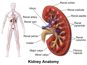
A frontal section through the kidney reveals an outer region called the renal cortex and an inner region called the medulla. The renal columns are connective tissue extensions that radiate downward from the cortex through the medulla to separate the most characteristic features of the medulla, the renal pyramids and renal papillae. The papillae are bundles of collecting ducts that transport urine made by nephrons to the calyxes of the kidney for excretion. The renal columns also divide the kidney into six to eight lobes and provide a supportive framework for vessels that enter and exit the cortex. The pyramids and renal columns taken together constitute the kidney lobes. Fig. 2.2 shows all these parts of the kidney. The video below provides an overview of the kidneys and the functions they provide in the human body.
(TED-Ed, 2015)
The renal hilum is the entry and exit site for the structures that service the kidneys—the vessels, nerves, lymphatics, and ureters. Emerging from the hilum is the renal pelvis, which is formed from the major and minor calyxes in the kidney. The smooth muscle in the renal pelvis uses peristalsis to funnel urine into the ureter.
Cortex
In a dissected kidney, it is easy to identify the cortex—it appears lighter in colour compared to the rest of the kidney. All the renal corpuscles as well as both the proximal convoluted tubules (PCTs) and distal convoluted tubules (DCTs) are found here.
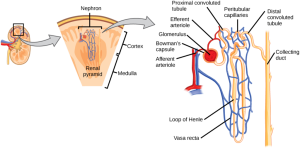
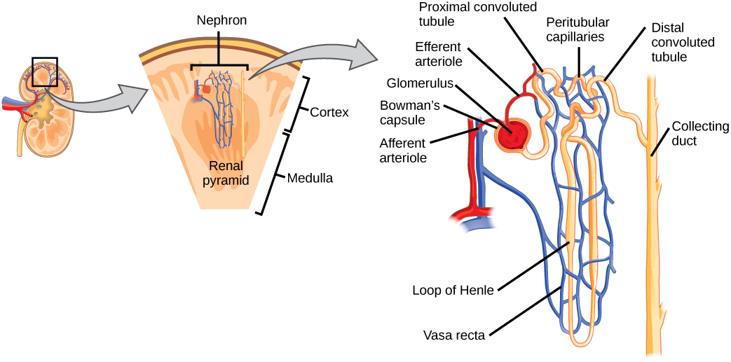
The urinary system’s ability to filter the blood resides in about 2 to 3 million tufts of specialized capillaries, called the glomeruli, which are distributed more or less equally between the two kidneys. Because the glomeruli filter the blood based mostly on particle size, large elements such as blood cells, platelets, antibodies, and albumen are excluded. The glomerulus is the first part of the nephron, which then continues as a highly specialized tubular structure responsible for creating the final urine composition.
Nephrons
Nephrons, as can be seen in Fig. 2.3, are the functional units of the kidney; they cleanse the blood and balance the constituents of circulation. The afferent arteriole forms a tuft of high-pressure capillaries called the glomerulus. The rest of the nephron consists of a continuous sophisticated tubule whose proximal end surrounds the glomerulus. This end is referred to as Bowman’s capsule. The glomerulus and Bowman’s capsule together form the renal corpuscle. The glomerular capillaries filter blood based on particle size. After passing through the renal corpuscle, the capillaries form a second arteriole, the efferent arteriole. Next, they form a capillary network around the more distal portions of the nephron tubule, the peritubular capillaries and the vasa recta, before returning to the venous system. As the glomerular filtrate progresses through the nephrons, these capillary networks recover most of the solutes and water, and return them to the circulation.
With up to 180 litres per day passing through the nephrons of the kidney, it is quite obvious that most of that fluid and its contents must be reabsorbed. That recovery occurs in the proximal convoluted tubule (PCT), loop of Henle, distal convoluted tubule (DCT), and the collecting ducts. Various portions of the nephron differ in their capacity to reabsorb water and specific solutes, which means that substances such as sodium (Na), potassium (K), chloride (Cl), urea, and many others are only absorbed, or reabsorbed, in certain parts of the nephron.
Urine Volumes
The table below is a great review of key terms in reference to urine volumes and gives a general idea of normal daily urine volumes as compared to what is seen when certain pathologies are present.
Table. 2.1. Urine Volumes
| Volume condition | Volume | Causes |
| Normal | 1–2 L/day | |
| Polyuria | >2.5 L/day | Diabetes mellitus, diabetes insipidus, excess caffeine or alcohol, kidney disease, sickle cell anemia, excessive water intake, and certain drugs, such as diuretics |
| Oliguria | 300–500 mL/day | Dehydration, blood loss, diarrhea, cardiogenic shock, kidney disease, and enlarged prostate |
| Anuria | <50 mL/day | Kidney failure, enlarged prostate, and obstruction, such as a kidney stone or tumour |
(Betts el al., 2013)
Attribution
Unless otherwise indicated, material on this page has been adapted from the following resource:
Betts, J. G., Young, K. A., Wise, J. A., Johnson, E., Poe, B., Kruse, D. H., Korol, O., Johnson, J. E., Womble, M., & DeSaix, P. (2013). Anatomy and physiology. OpenStax. https://openstax.org/details/books/anatomy-and-physiology, licensed under CC BY 4.0
References
Ernstmeyer, K., & Christman, E. (Eds.). (2020). Nursing pharmacology. Chippewa Valley Technical College. https://wtcs.pressbooks.pub/pharmacology/, licensed under CC BY 4.0
TED-Ed. (2015, February 9). How do your kidneys work? – Emma Bryce [Video]. YouTube. https://www.youtube.com/watch?v=FN3MFhYPWWo
Image Credits (images are listed in order of appearance)
Urinary System (Female) by BruceBlaus, CC BY-SA 4.0
Blausen 0592 KidneyAnatomy 01 by BruceBlaus, CC BY-SA 3.0
Figure 41 03 03 by CNX OpenStax, CC BY-SA 4.0
the contraction and relaxation of muscles that move in a wave down a tube, for example, in the intestines

.png)