
9.2 The Nervous System
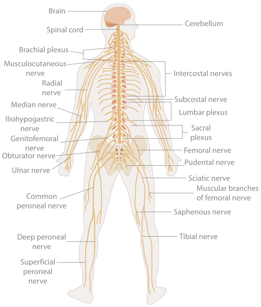
The nervous system, shown in Fig. 9.1, is a very complex system and is responsible for controlling much of the body, including both voluntary and involuntary functions. It receives information about the environment around us, then creates responses to that information. This system is also responsible for taking sensory input and integrating it with other sensations, memories, emotional states, and learning. The nervous system can be divided into two main components: the central nervous system and the peripheral nervous system. From there, it is subdivided even further by functions and components.
Components of the Nervous System
As mentioned above, the two main components of the. nervous system are the central nervous system (CNS) and the peripheral nervous system (PNS).
Central nervous system (CNS): The brain and the spinal cord make up the central nervous system. The brain is described in terms of its major regions, which include the cerebrum, diencephalon, brain stem, and cerebellum. The regulation of homeostasis and conscious experiences are controlled in the brain. Reflexes and the integration of sensory and motor pathways are handled in the spinal cord.
Peripheral nervous system (PNS): This part of the nervous system connects the central nervous system with the rest of the body. The nerves, axons, and ganglia that make up the PNS are found throughout the body in other organs and even in other systems, such as the digestive system, as well as the eyes, ears, nose, and various other locations. Messages travel back and forth from the CNS to the muscles, organs, and senses in peripheral areas of the body. When sensory neurons carry messages and various forms of sensory information towards the CNS, they are considered afferent fibres. When the CNS uses motor neurons to carry instructions from the CNS to the muscles, they are called efferent fibres. Messages constantly travel back and forth along neurons between the CNS and the periphery.
The PNS is subdivided into two components: the somatic nervous system and the autonomic nervous system.
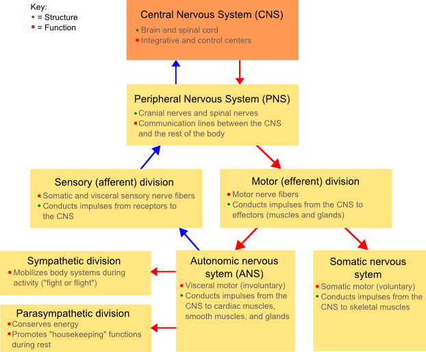
Somatic nervous system: This part of the PNS is responsible for conscious perception of the environment and for voluntary responses to that perception through the use of skeletal muscles.
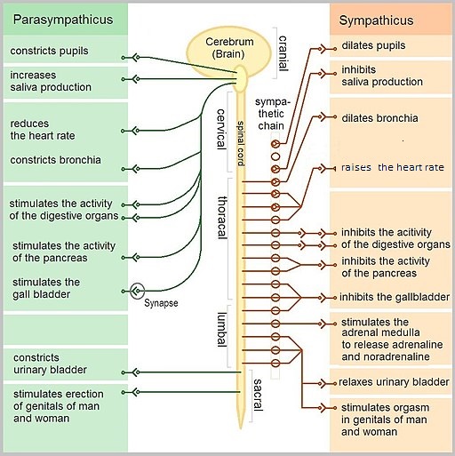
Autonomic nervous system: This part of the PNS handles involuntary responses that the brain controls without the need for conscious thought. It consists of the sympathetic and parasympathetic nervous systems and uses a balance of the two to regulate the body’s involuntary functions, including heart rate, respiratory rate, digestion, and sweating:
-
- Sympathetic nervous system: Associated with the fight-or-flight response
- Parasympathetic nervous systems: Focuses on what could be called “rest and digest”
, and Fig. 9.3 discusses the differences between the parasympathetic and sympathetic nervous systems.
Neurons
Neurons are the cells considered to be the basis of nervous tissue within the body (Betts et al., 2013). They are responsible for the electrical signals that communicate information about sensations and produce movements in response to those stimuli, along with starting thought processes in the brain. An important part of the function of neurons is in their unique structure. The shape and the parts of these cells are what make the numerous connections within the nervous system possible (Betts et al., 2013).
Parts of a Neuron
The main part of a neuron is the cell body, which contains the nucleus and most of the major organelles (Betts et al., 2013). The axon is a fibre that emerges from the cell body and projects to target cells. A single axon can branch repeatedly to communicate with many target cells (Betts et al., 2013). It is the axon that continues the nerve impulse, or electrical signal, which is then communicated to one or more cells. The neurons also have dendrites, which receive information from other neurons at specialized areas of contact called synapses. The dendrites are usually highly branched, providing locations for other neurons to communicate with the cell body. Information flows through a neuron from the dendrites, across the cell body, and down the axon (Betts et al., 2013). Fig. 9.4 provides a diagram of all the main components of a neuron.
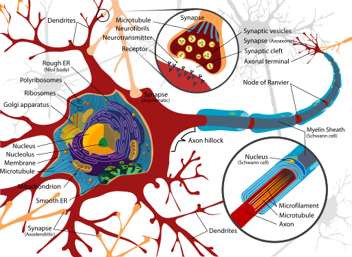
Axons are wrapped by an insulating substance called myelin, which is made from glial cells (Betts et al., 2013). Myelin acts as insulation but there are also gaps in the myelin covering of an axon (Betts et al., 2013). Each gap is called a node of Ranvier and is important to the way electrical signals travel down the axon. At the end of the axon is the axon terminal, where there are usually several branches extending toward the target cells; each branch ends in an enlargement called a synaptic end bulb. These bulbs are what make the connections with the target cells at the synapse (Betts et al., 2013). The axon terminal is also where the electrical signals are changed to chemical signals called neurotransmitters, which are used to communicate with the next group of nerve cells. The video below provides a quick overview of the neuron and how it functions.
(Neuroscientifically Challenged, 2014)
Communication in the Nervous System
Understanding how communication occurs within the nervous system will help you understand the mechanism of action of medication that works by influencing the neurotransmitters. There are two types of connections between electrically active cells: chemical synapses and electrical synapses. In an electrical synapse, there is a direct connection between two cells so that ions can pass from one cell to the next. In a chemical synapse, electrical impulses signal the release of a chemical signal, a neurotransmitter, that then travels to, and binds with, a target cell. We will be focusing on the communication of a neurotransmitter in a chemical synapse because most of the medications discussed affect this form of connection.
Once in the synaptic cleft, the space between two neurons, the neurotransmitter diffuses the short distance to the postsynaptic membrane and can interact with neurotransmitter receptors (Fig. 9.5). Receptors are specific for the neurotransmitter, and the two fit together like a key and lock. A neurotransmitter binds to its receptor and will not bind to receptors for other neurotransmitters, making the binding a specific chemical event.
When the neurotransmitter binds to the receptor, the cell membrane of the target neuron changes its electrical state, and a new, graded potential begins. If that graded potential is strong enough to reach threshold, the next neuron generates an action potential and continues the message, or electrical signal. This continues over and over until the message reaches its destination in the brain.
Neurotransmitters
Neurotransmitters are chemical messengers that our body cannot work without. An important thing to remember about neurotransmitters and signalling chemicals is that the effect is entirely dependent on the receptor. There are a number of different types of neurotransmitters in the body:
- Dopamine
- Serotonin
- Histamine
- Norepinephrine
- Epinephrine
- Glutamate
- Acetylcholine
Examples of Neurotransmitter Functions
Any alteration in central nervous system (CNS) function can be related to abnormal impulse transmission and can result from an imbalance of neurotransmitters. A person with an imbalance of neurotransmitters may have the signs and symptoms of a CNS disorder or pathology. The medications used to treat CNS disorders do so by mimicking or blocking the neurotransmitter based on the imbalance caused by the condition. Medications can also be used to either stimulate or depress the effect of the neurotransmitter. For example, CNS depressants alter the brain by decreasing the excitability of neurotransmitters, blocking their receptor site or increasing the inhibitory neurotransmitter. Conversely, CNS stimulants increase brain activity by increasing the excitability of neurotransmitters, decreasing the inhibitory neurotransmitters or blocking their receptor sites.
Examples:
- Norepinephrine is often associated with the fight-or-flight response. Abnormal levels of this neurotransmitter are also associated with depression and decreased alertness and interest, along with possible palpitations, anxiety, and panic attacks.
- Dopamine is strongly linked to motor movement and cognition. It influences movement and can be associated with ADHD, paranoia, and schizophrenia.
- Serotonin is heavily involved in many bodily processes. Abnormal levels of serotonin can affect sleep, libido, mood, and temperature regulation. Alterations of this neurotransmitter have been linked to many mental health issues such as depression, bipolar disorder, anxiety, and body disorders.
Attribution
Unless otherwise indicated, material on this page has been adapted from the following resource:
Ernstmeyer, K., & Christman, E. (Eds.). (2020). Nursing pharmacology. Chippewa Valley Technical College. https://wtcs.pressbooks.pub/pharmacology/ licensed under CC BY 4.0
References
Betts, J. G., Young, K. A., Wise, J. A., Johnson, E., Poe, B., Kruse, D. H., Korol, O., Johnson, J. E., Womble, M., & DeSaix, P. (2013). Anatomy and physiology. OpenStax. https://openstax.org/details/books/anatomy-and-physiology licensed under CC BY 4.0
Neuroscientifically Challenged. (2014, July 22). 2-minute neuroscience: The neuron [Video]. YouTube. https://www.youtube.com/watch?v=6qS83wD29PY
Image Credits (images are listed in order of appearance)
TE-Nervous_system_diagram by The Emirr, CC BY 3.0
NSdiagram by Fuzzform, CC BY-SA 3.0
The_Autonomic_Nervous_System by Geo-Science-International, CC0 1.0
Complete_neuron_cell_diagram_en by LadyofHats, Public domain
Generic_Neurotransmitter_System by National Institute on Drug Abuse (NIDA), Public domain
a membrane-enclosed organelle within a cell that contains the chromosomes
small structures in a cell that are surrounded by a membrane and have a specific function
the point of contact between neurons where information is passed from one neuron to the next
a type of cell that provides physical and chemical support to neurons and maintains their environment
