
6.9 Nervous System
Overview and Functions
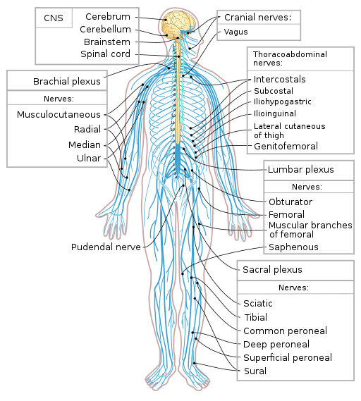
The nervous system (Fig. 6.24) is a very complex system and is responsible for controlling much of the body, including both voluntary and involuntary functions. It receives information about the environment around us, and then creates responses to that information. This system is also responsible for taking sensory input and integrating it with other sensations, memories, emotional states, and learning. The nervous system can be divided into two main components: the central nervous system and the peripheral nervous system. From there, it is further subdivided by functions and components.
(CrashCourse, 2015)
Components of the Nervous System
The two main components of the nervous system are the central nervous system (CNS) and the peripheral nervous system (PNS).
Central nervous system (CNS): The brain and the spinal cord make up the central nervous system. The brain is described in terms of its major regions, which include the cerebrum, diencephalon, brain stem, and cerebellum. The regulation of homeostasis and conscious experiences are controlled in the brain. Reflexes and the integration of sensory and motor pathways are handled in the spinal cord.
Peripheral nervous system (PNS): This part of the nervous system connects the central nervous system with the rest of the body. The nerves, axons, and ganglia that make up the PNS are found throughout the body. Many are found in other organs and even in other systems, such as the digestive system, as well as the eyes, ears, nose, and various other locations. Messages travel back and forth from the CNS to the muscles, organs, and senses in the peripheral areas of the body. When sensory neurons carry messages and various forms of sensory information towards the CNS, they are considered afferent fibres. When the CNS uses motor neurons to carry instructions from the CNS to the muscles, they are called efferent fibres. Messages continually go back and forth along neurons between the CNS and the periphery. The PNS has two subdivisions as well—the somatic nervous system and the autonomic nervous system. Fig. 6.25 provides an overall picture of the nervous system and its components.
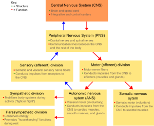
Somatic nervous system: This part of the PNS is responsible for conscious perception of the environment and for voluntary responses to that perception through use of skeletal muscles.
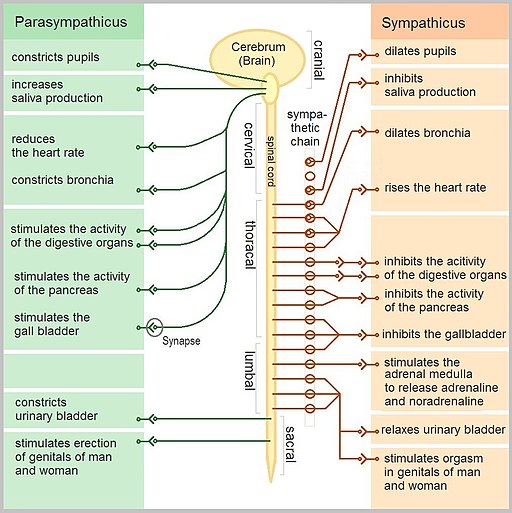
Autonomic nervous system: This part of the PNS handles involuntary responses that the brain controls without the need for conscious thought. It consists of the sympathetic and parasympathetic nervous systems and uses a balance of the two to regulate the body’s involuntary functions, including heart rate, respiratory rate, digestion, and sweating:
- Sympathetic nervous system: Associated with the fight-or-flight response
- Parasympathetic nervous systems: Focuses on what could be called “rest and digest”
Fig. 6.26 shows how the sympathetic and parasympathetic nervous systems work within the body. Both affect the same areas of the body but in a different manner.
Combining Forms
Table 6.8. Combining Forms
| COMBINING FORM | MEANING | EXAMPLE OF USE IN MEDICAL TERMS |
|---|---|---|
| aur/o | ear | aural hematoma |
| cerebell/o | cerebellum | cerebellitis |
| cerebr/o | cerebrum | cerebral |
| dur/o | dura mater | durotomy |
| encephal/o | brain | encephalopathy |
| medull/o | medulla oblongata | medullary |
| mening/o | meninges | meningitis |
| myel/o | spinal cord | myelitis |
| myring/o | eardrum | myringoplasty |
| neur/o | nerve | neurologist |
| ocul/o | eye | ocular |
| ophthalm/o | eye | ophthalmologist |
| optic/o | eye | optical |
| ot/o | ear | otalgia |
| phak/o | lens of the eye | aphakia |
| retin/o | retina | retinopathy |
| tympan/o | eardrum | tympanostomy |
Common Pathologies
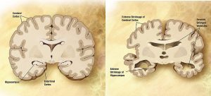
Alzheimer’s disease: This form of dementia is characterized by the accumulation of beta-amyloid plaque, a type of dense protein found in the cerebral cortex. It is a degenerative disease in which individuals experience memory loss and confusion. As shown in Fig. 6.27, the brain atrophies (shrinks) as the condition progresses.
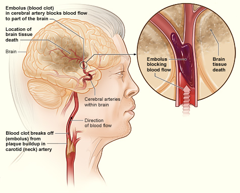
Cerebrovascular accident (CVA): Commonly known as a stroke, this condition is characterized by loss of blood flow to a part of the brain (Fig. 6.28). There are two types of strokes: ischemic and hemorrhagic. An ischemic stroke is caused by a blocked or narrowed vessel in the brain that obstructs blood flow. The cause is often a blood clot or fatty deposit. In a hemorrhagic stroke, bleeding to the brain occurs because of a damaged blood vessel.
Key Concept
The mnemonic FAST helps people remember what to look for when a cerebrovascular accident (stroke) is suspected:
- Look at the person’s face. Do they have problems moving Face muscles and making regular facial expressions?
- Ask the person to raise their Arms above their head. Can the person lift one arm but not the other?
- Has the person’s Speech changed? Are they slurring words or having trouble saying things?
- If any of these things have happened, then it is Time to call for help.
Cataract: This condition clouds the normally clear lens of the eye. A possible cause is a decrease in the flexibility of the eye lens because of the aging process; also, some infants are born with congenital cataracts (Carter & Rutherford, 2020).
Concussion: A concussion is a traumatic injury to the brain from an impact (Fig. 6.29). Symptoms include memory loss, headaches, and difficulty concentrating. There may or may not be a loss of consciousness (Ernstmeyer & Christman, 2020).

Epilepsy: This is a chronic condition of reoccurring seizures. Symptoms of a seizure include muscle rigidity, jerking, muscle twitching, and muscle weakness. Epilepsy is often diagnosed with the use of an electroencephalogram, and then treated with a combination of a few different medications (Ernstmeyer & Christman, 2020).
Glaucoma: This condition is an increase in intraocular pressure owing to an increase in fluid build-up in the anterior compartment of the eye.
Hemiplegia: This is paralysis of one side of the body and is often the result of a stroke.
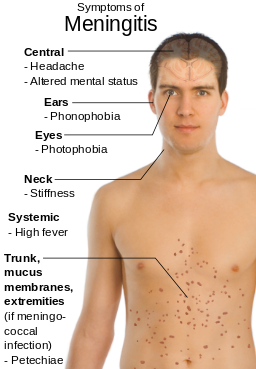
Meningitis: This disease is inflammation of the meninges, which are the membranes around the CNS. It can be caused by a virus or bacteria. Symptoms include fever, chills, nausea, vomiting, neck soreness, confusion, and severe headaches.
Multiple sclerosis (MS): This is an autoimmune disease in which antibodies produced by the lymphocytes attack the myelin (the insulating sheath around the nerves) in the nervous system. As a result, the myelin becomes inflamed, destroyed, or scarred. Myelin is needed to maintain nerve condition, and damage to it results in nerve conduction in the CNS to be slowed. Symptoms of MS include vision issues, numbness and tingling in the extremities, pain, muscle spasms, issues with balance, and muscle weakness.
Paraplegia: This is paralysis of the lower half of the body and is often the result of trauma.
Parkinson’s disease: This progressive disease of the nervous system affects the ability to move. Onset usually occurs in middle adulthood, and symptoms worsen over time. There is no cure, and the cause is unknown. Symptoms include tremors, muscle rigidity, unstable posture, issues with gait, and slow body movements (Ernstmeyer & Christman, 2020).
Syncope: Another term for this condition is fainting. It is characterized by sudden, temporary loss of consciousness.
Tinnitus: This condition is characterized by ringing in the ears. It is often caused by inflammation of the middle ear or exposure to loud noise (Ernstmeyer & Christman, 2020).
Transient ischemic attack (TIA): A TIA is similar to a stroke but does not last as long. If the classic symptoms of a stroke resolve without treatment, a TIA is possibly the cause.
Exercise
Attribution
Unless otherwise indicated, material on this page has been adapted from the following resource:
Betts, J. G., Young, K. A., Wise, J. A., Johnson, E., Poe, B., Kruse, D. H., Korol, O., Johnson, J. E., Womble, M., & DeSaix, P. (2013). Anatomy and physiology. OpenStax. https://openstax.org/details/books/anatomy-and-physiology licensed under CC BY 4.0
References
Carter, K., & Rutherford, M. (2020). Building a medical terminology foundation. eCampusOntario. https://ecampusontario.pressbooks.pub/medicalterminology/ licensed under CC BY 4.0
CrashCourse. (2015, February 23). The nervous system, part I: Crash Course A & P #8 [Video]. YouTube. https://www.youtube.com/watch?v=qPix_X-9t7E&list=PL8dPuuaLjXtOAKed_MxxWBNaPno5h3Zs8&index=9
Ernstmeyer, K., & Christman, E. (Eds.). (2020). Nursing pharmacology. Chippewa Valley Technical College. https://wtcs.pressbooks.pub/pharmacology/ licensed under CC BY 4.0
Image Credits (images are listed in order of appearance)
Nervous system diagram-en by Medium69 and Jmarchn, CC BY-SA 4.0
NSdiagram by Fuzzform, CC BY 3.0
The Autonomic Nervous System by Geo-Science-International, CC0 1.0
Alzheimer’s disease brain comparison by Alzheimer’s Disease Education and Referral (ADEAR) Center, National Institute on Aging, Public domain
Stroke ischemic by National Heart, Lung, and Blood Institute, Public domain
Concussion Anatomy by Max Andrews, CC BY 3.0
Meningitis by Mikael Häggström, Public domain
A mass of blood within the ear
Inflammation of the cerebellum
Pertaining to the cerebrum
An incision into the dura mater
A disease or disorder of the brain
Pertaining to the medulla oblongata
Inflammation of the meninges
Inflammation of the spinal cord
Surgical correction of the eardrum
A specialist in the study of nerves (nervous system)
Pertaining to the eye
A specialist in the study of the eye
Pertaining to the eye
The condition of pain in the ear
The condition of no lens of the eye
A disease condition of the retina
A small tube inserted into the eardrum
