
6.11 Digestive System
Overview and Functions
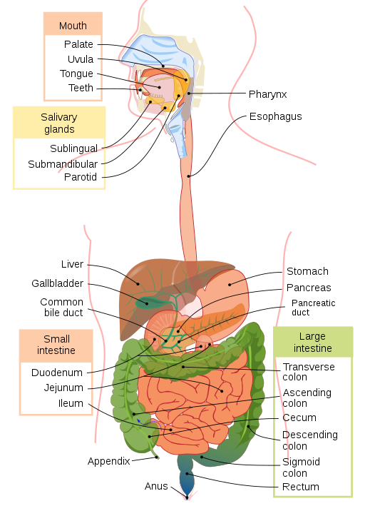
The function of the digestive system (Fig. 6.34) is to break down food, release its nutrients, and absorb those nutrients into the body. The small intestine is the pillar of the system. It is where the majority of digestion occurs and where most of the released nutrients are absorbed into the blood. However, each of the digestive system organs makes a vital, and necessary, contribution to this process.
(CrashCourse, 2015)
Components of the Digestive System
Mouth: The mouth begins the digestive process by ingesting food and chewing it, starting the chemical breakdown of carbohydrates, then moving the food into the pharynx.
Salivary glands: These small exocrine glands are located within the mouth and tongue. They constantly produce saliva, and on average, secrete about 1 to 1.5 L of saliva each day.
Teeth: Similar to bones, the teeth are used to tear, grind, and otherwise mechanically break down food.
Pharynx: This organ is also known as the throat and is involved in both digestion and respiration. When food enters the pharynx, involuntary muscle contractions close off the airways to prevent food from entering. When a person is not eating, the airways are open for air to enter from the mouth and nasal cavities.
Esophagus: This muscular tube connects the pharynx to the stomach. It is approximately 25 cm (10 in) long and is collapsed when not engaged in swallowing.
Stomach: The stomach participates in almost all the digestive activities except for ingestion and defecation. There are four main regions to the stomach: cardia, fundus, body, and pylorus.
Small intestine: Almost all digestion occurs in the small intestine, and practically all absorption takes place there as well. The small intestine has three parts: duodenum, jejunum, and ileum.
Key Concept
The small intestine is about 3 m (10 ft) long in a living person, but about twice as long in a cadaver as a result of the loss of muscle tone. This also makes it about five times longer than the large intestine.
Large intestine: The primary function of the large intestine is to complete the absorption of nutrients and water, synthesize vitamins, form feces, and eliminate feces from the body. The large intestine has four main regions: cecum, colon, rectum, and anus.
Liver: The largest gland in the body, the liver weighs approximately 1.4 kg (3 lb) in an adult. It is also one of the most important organs because of its role as an accessory digestive organ and in metabolism and regulation.
Pancreas: The functions of the pancreas involve a mix of exocrine functions, such as secreting digestive enzymes, and endocrine functions, such as releasing hormones into the blood.
Gallbladder: This organ is approximately 8 to 10 cm (3 to 4 in ) long. It stores, concentrates, and then sends bile into the duodenum (part of the small intestine).
Combining Forms
Table 6.10. Combining Forms
| COMBINING FORM | MEANING | EXAMPLE OF USE IN MEDICAL TERMS |
|---|---|---|
| abdomin/o | abdomen | abdominal |
| an/o | anus | anal |
| append/o | appendix | appendectomy |
| appendic/o | appendix | appendicitis |
| cholecyst/o | gallbladder | cholecystostomy |
| col/o | colon | colectomy |
| colon/o | colon | colonography |
| duoden/o | duodenum | duodenoscope |
| enter/o | intestine | gastroenteritis |
| esophag/o | esophagus | esophagogastrectomy |
| gastr/o | stomach | hemigastrectomy |
| hepat/o | liver | hepatitis |
| ile/o | ileum | ileostomy |
| jejun/o | jejunum | jejunoileitis |
| lapar/o | abdomen | laparotomy |
| or/o | mouth | oral |
| pancreat/o | pancreas | pancreatic |
| pharyng/o | pharynx | pharyngitis |
| proct/o | anus and rectum | proctologist |
| rect/o | rectum | rectal |
| sigmoid/o | sigmoid colon | sigmoidoscopy |
| stomat/o | mouth | stomatologist |
Common Pathologies
Celiac disease: This is an immune sensitivity reaction that occurs in the small intestine when gluten is consumed. Damage to the small intestine will occur if gluten consumption continues (Carter & Rutherford, 2020).
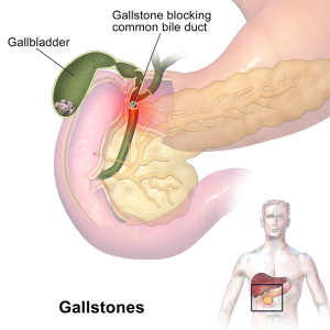
Cholecystitis: This condition is usually caused by gallstones that develop and block the bile duct. As a result, the gallbladder becomes inflamed.
Cholelithiasis: This condition is also known as gallstones (Fig. 6.25); the stones are pieces of solid material that form in the gallbladder (WebMD, 2022a). Gallstones can range in size from a grain of rice to a golf ball, and as they get larger, the chance that they will block the bile duct increases. When a blockage occurs, the result is pain and discomfort in the gallbladder region.
Cirrhosis: This condition is characterized by liver scarring. Cirrhosis is a chronic condition that cannot be cured, and as it progresses, it can become life threatening. Chronic alcoholism and some liver diseases can cause cirrhosis (Mayo Clinic, 2022).
Colon cancer: One of the most malignant types of cancer, smoking, excessive alcohol intake, a diet high in animal fats, and a family history of colon cancer increase a person’s chances of being diagnosed. Common symptoms include constipation, diarrhea, cramping, abdominal pain, and bleeding from the rectum.
Colonic polyposis: Also known a polyps, this condition is characterized by small growths that protrude outward from the intestinal wall. Most are benign, but some can become cancerous. Usually this condition is only present in those over the age of 50 (Carter & Rutherford, 2020).
Diverticulitis: This condition is caused by inflammation of the diverticula (see Diverticulosis, below) (WebMD, 2022b). Symptoms include severe abdominal pain and fever.
Diverticulosis: This condition is characterized by small bulging pockets, known as diverticula, that form within the walls of the digestive tract (WebMD, 2022b). Often there are no signs or symptoms unless they become inflamed. The pockets can grow in size and food can become stuck, resulting in diverticulitis (WebMD, 2022b).
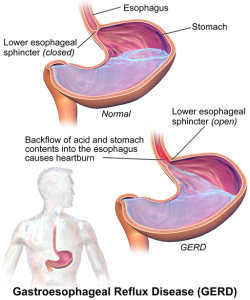
Gastroesophageal reflux disease (GERD): This condition occurs when the lower esophageal sphincter does not fully close, and the contents of the stomach travel back up into the esophagus (Fig. 6.36). The backwash of stomach acid, known as acid reflux, irritates the lining of the esophagus, causing heartburn, chest pain, and difficulty swallowing.
Hepatitis A, B, and C: Hepatitis is inflammation of the liver and can be caused by various factors. Viruses, alcohol consumption, toxins, medications, and even autoimmune responses can result in hepatitis (Carter & Rutherford, 2020).
Hernia: This condition occurs when fatty tissue or an organ pushes through a weak area of the surrounding muscle or connective tissues. The most common hernias occur in the abdomen or groin areas.
Inflammatory bowel disease (IBD): This disease occurs when a part of the digestive tract becomes inflamed. The two most common types of IBD are Crohn’s disease, which occurs in the upper GI tract, and ulcerative colitis, which occurs in the colon. These are chronic conditions that can be treated but not cured (Carter & Rutherford, 2020).
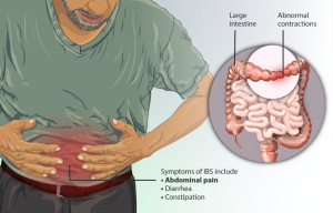
Irritable bowel syndrome (IBS): The cause of IBS is unknown. Symptoms include bloating, gas, and abdominal pain (Carter & Rutherford, 2020). Usually those that suffer from IBS are advised to make dietary and lifestyle changes to deal with the symptoms they experience.
Jaundice: This condition is caused when bilirubin, a yellowish pigment produced during the normal breakdown of red blood cells, increases because it cannot be removed effectively from circulation. Often a failing liver causes this condition, giving a yellowish tinge to the skin, eyes, and mucosa. Fig. 6.38 below shows a person exhibiting the common symptoms of jaundice.

Ulcers: These sores in the lining of the stomach are caused by gastric juices breaking down the stomach mucosa. This causes erosions of the stomach lining, which can heal on their own, but deeper and larger ones become ulcers. Certain medications, such as non-steroidal anti-inflammatory drugs (NSAIDS), and infections, like Helicobacter pylori, can contribute to ulcer formation.
Exercise
Attribution
Unless otherwise indicated, material on this page has been adapted from the following resource:
Betts, J. G., Young, K. A., Wise, J. A., Johnson, E., Poe, B., Kruse, D. H., Korol, O., Johnson, J. E., Womble, M., & DeSaix, P. (2013). Anatomy and physiology. OpenStax. https://openstax.org/details/books/anatomy-and-physiology licensed under CC BY 4.0
References
Carter, K., & Rutherford, M. (2020). Building a medical terminology foundation. eCampusOntario. https://ecampusontario.pressbooks.pub/medicalterminology/ licensed under CC BY 4.0
CrashCourse. (2015, September 7). The digestive system, part 1: Crash Course A&P #33 [Video]. YouTube. https://www.youtube.com/watch?v=yIoTRGfcMqM&list=PL8dPuuaLjXtOAKed_MxxWBNaPno5h3Zs8&index=34
Ernstmeyer, K., & Christman, E. (Eds.). (2020). Nursing pharmacology. Chippewa Valley Technical College. https://wtcs.pressbooks.pub/pharmacology/ licensed under CC BY 4.0
Mayo Clinic. (2022). Cirrhosis. https://www.mayoclinic.org/diseases-conditions/cirrhosis/symptoms-causes/syc-20351487
WebMD. (2022a). Gallstones. https://www.webmd.com/digestive-disorders/gallstones
WebMD. (2022b). What is diverticulosis? https://www.webmd.com/digestive-disorders/what-is-diverticulosis
Image Credits (images are listed in order of appearance)
Digestive system diagram by Mariana Ruiz and Jmarchn, Public domain
Gallstones by BruceBlaus, CC BY-SA 4.0
GERD by BruceBlaus, CC BY-SA 4.0
Depiction of a person suffering from Irritable Bowel Syndrome by myUpchar, CC BY-SA 4.0
Scleral Icterus by Sheila J. Toro, CC BY 4.0
Pertaining to the abdomen
Pertaining to the anus
Removal of the appendix
Inflammation of the appendix
An opening into the gallbladder; often done for drainage
Removal of part of the colon
The processing of a recording (often a CT scan) of the colon
An instrument used to view the duodenum
Inflammation of the stomach and intestines
Removal of part of the esophagus and stomach
Removal of half the stomach
Inflammation of the liver
An opening (stoma) in the ileum
Inflammation of the jejunum and ileum (a type of Crohn's disease)
Cutting into the abdomen
Pertaining to the mouth
Pertaining to the pancreas
Inflammation of the pharynx
A specialist in the study of the anus and rectum
Pertaining to the rectum
The process of viewing the sigmoid colon
A specialist in the study of the mouth

.png)