
8.6 Clinical Tests and Procedures
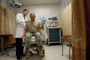
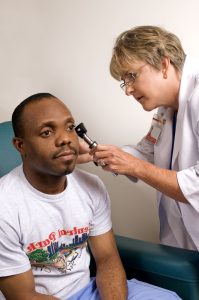
A vast number of clinical tests and procedures are carried out in medical settings every day. This section will focus on the most common ones done in a hospital on a day-to-day basis and are used, in some way, for diagnostic purposes. Although a number of other clinical procedures can be performed, especially on more specialized units in a hospital, those discussed below are a good representation of the clinical tests and procedures you will likely see in your future medical career. Fig. 8.66 and Fig. 8.67 show doctors performing physical examinations on patients.

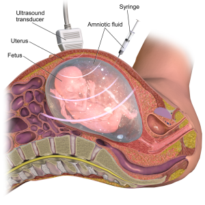
Amniocentesis: In this procedure, a physician takes a sample of amniotic fluid from a pregnant woman. Not all pregnancies require this test; it is performed to determine whether the fetus has any chromosomal disorders. Amniocentesis may be suggested as an option for a patient if such disorders are suspected or if the patient is over a certain age because that can increase the risk that the fetus may present with a chromosomal disorder. The physician uses a syringe guided by an ultrasound, as shown in Fig. 8.68, to take a sample of amniotic fluid, which is then sent to the lab for analysis.
Arthrocentesis: In this test, a syringe is used to remove fluid from a joint. Possible reasons for this procedure would be to test the fluid or to remove fluid that may be causing the patient discomfort (Carter & Rutherford, 2020).
Aspiration: This medical procedure involves the removal of something from the body (NLM, 2022). The substance may vary; however, it could potentially be body fluids, part of a bone, or air. Aspiration is used when a physician performs a biopsy and is the act of using a needle to withdraw a sample to be sent to the lab.
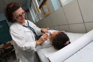
Auscultation: This test involves placing a stethoscope over a part of the body, most often the lungs, heart, or abdomen, and listening for either the lack of sounds or appropriate sounds for that area. Heart sounds can be heard with a classic stethoscope, but with a cardiac stethoscope, as shown in Fig. 8.69, it is also possible to hear the opening and closing of valves within the heart. Listening to lung sounds can determine whether there is equal air entry to both lungs and whether or not fluid if present. Finally, the abdomen can be assessed to determine whether there are bowel sounds, or a lack thereof, which could indicate the absence of movement through the gastrointestinal tract.
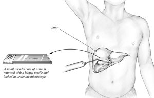
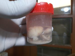
Biopsy (Bx): This procedure involves taking a sample of material from the body and sending it to the lab for analysis. Biopsies are often completed to determine whether a growth is cancerous or benign. Fig. 8.7 explains how a liver biopsy is performed, and Fig. 8.71 shows a sample obtained from a biopsy.
Key Concept
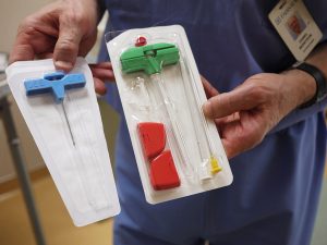
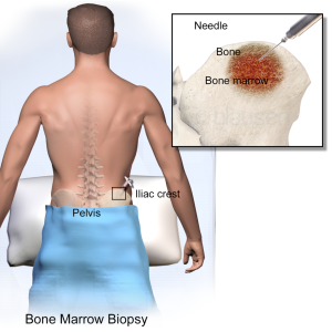
A bone marrow biopsy is a specific type of biopsy that involves taking a sample of bone marrow (Carter & Rutherford, 2020). Often, this is used in the diagnosis and treatment of various severe forms of anemia, such as thalassemia major and sickle cell anemia, as well as some types of cancer, specifically leukemia. The instruments used to obtain a bone marrow sample are shown in Fig. 8.72, and Fig. 8.73 shows the procedure and the area of the body that the sample is typically taken from.
Bronchoscopy: This procedure involves visualization of trachea and bronchi with a bronchoscope. Some common reasons for a bronchoscopy include chronic cough, infection, or an anomaly on a chest X-ray or other scan (Johns Hopkins, 2022). Bronchoscopies can also be used to take samples or remove foreign objects from the airway (Carter & Rutherford, 2020).
Colonoscopy: In this procedure, a colonoscope is used to visualize the large intestine and rectum. A colonoscopy may be recommended for patients who have unexplained weight loss, abdominal pain, changes to bowel movements, or bleeding from the anus (NIDDK, 2017). This procedure can also be beneficial for cancer and polyp screening.
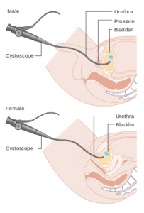
Cystoscopy: This procedure is completed with a cystoscope and, as shown in Fig. 8.74, involves visualizing the bladder. The cystoscope is inserted via the urethra and passed into the bladder (NHS, 2022). This test is used to check for bladder and urethral issues. The images appear similar to what is seen in Fig. 8.75. Patients may have this procedure if they experience hematuria, recurrent urinary tract infections, pelvic pain, or difficulty urinating. Biopsy samples can also be taken during the cystoscopy, and bladder stones may also be removed (Carter & Rutherford, 2020).
Digital rectal exam (DRE): In this procedure, a physician palpates the prostate to check for masses or anomalies. This is often done in conjunction with a prostate-specific antigen (PSA) test to assess for prostate cancer (Carter & Rutherford, 2020).
Electrocardiogram (ECG): This procedure can be performed by various medical professionals, including technicians, nurses, or physicians. An ECG is a recording of the electrical activity of the heart. Electrodes are placed on the patient’s chest, arms, and legs to create the electrical reading. It only takes a few minutes for the electrocardiograph to produce the electrical record. This test is done to assess for myocardial infarctions, arrhythmias, and other heart issues.
Electroencephalogram (EEG): In an EEG, electrodes are applied to a patient’s scalp to measure electrical activity in the brain. This test is used to help diagnose seizures, psychiatric conditions, tumours, and other brain disorders such as inflammation and injuries (Carter & Rutherford, 2020).
Electromyography (EMG): This procedure assesses the function of nerve cells that control muscles. Electrodes can either be attached to the skin or inserted into muscles to record electrical impulses. An EMG can assist with the diagnosis of nerve issues in different muscles and peripheral nerves (Carter & Rutherford, 2020).
Gastroscopy: For this test, a physician places a flexible gastroscope through the patient’s mouth and passes it down their throat to the stomach (Cancer Research UK, 2022). This allows the physician to view the entire upper digestive tract and take biopsy samples, which can help in the treatment of conditions such as stomach ulcers (Carter & Rutherford, 2020).
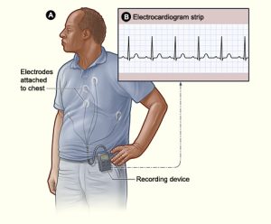
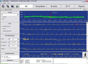
Holter monitor: This test is similar to an ECG, but the main difference is that the patient wears the Holter monitor for a 24-hour period (sometimes longer), and electrical readings are taken for the whole duration. This gives the physician a better idea of the patient’s cardiac electrical activity throughout their day, versus an ECG, which only takes readings for a short time. Fig. 8.76 shows a patient wearing a Holter monitor, and Fig. 8.77 is a sample of readings from a Holter monitor.
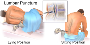
Lumbar puncture (LP): Also known as a spinal tap, this procedure involves withdrawing a sample of cerebrospinal fluid from the lower lumbar region, as shown in Fig. 8.78. The sample is taken from that location in the spine because the spinal cord ends in the upper lumbar region. Samples of cerebrospinal fluid are analyzed in the lab to diagnose, or rule out, diseases that affect the nervous system.
Ophthalmoscopy: This procedure can be completed by a physician or a specialist of the eye and involves visualization of the exterior and interior of a patient’s eye. It can assist with the diagnosis of eye injures and pathologies such as glaucoma and cataracts (Carter & Rutherford, 2020).
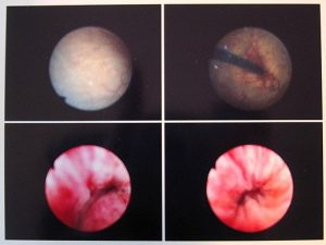
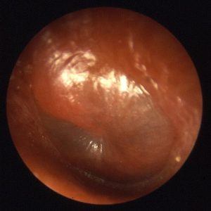
Otoscopy: Using an otoscope, a physician will visualize the inner ear, tympanic membrane, and ear canal during this assessment. Otoscopy may be done for various reasons, including pain or foreign objects in the ear, or pathologies such as infections that affect the ear (Carter & Rutherford, 2020). The procedure is shown in Fig. 8.79, and Fig. 8.80 is an example of what a physician may see when performing the procedure.
Palpation: Physicians, nurses, and other healthcare professionals use palpation in their assessment of patients. To perform the procedure, the healthcare professional uses their hands to assess for pain, tenderness, and distention. An abdominal assessment, for example, often involves palpating the four abdominal quadrants for bladder or bowel distention. Pain or tenderness could possibly indicate an inflammatory condition or bowel obstruction (Doyle & McCutcheon, 2020).
Pap smear: Also referred to as a pap test, this procedure involves collecting cells from the cervix. This is done to test for changes in the cells that may indicate cancer or to assess whether cancer may develop in the future (Mayo Clinic, 2022a). A pap smear should be part of regular health screening for women, and the interval between tests varies depending on age and prior medical history.
Percussion: This is similar to palpation in that it involves a physical assessment of the patient using the hands. In this case, however, it involves tapping, which produces different sounds and this helps determine if the underlying tissues are filled with air, fluid or a solid tissue. For example, over the abdomen, to assess for rigidity and pain (Doyle & McCutcheon, 2020).
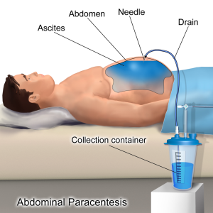
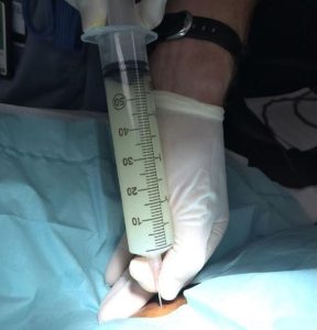
Paracentesis: This procedure uses a hollow needle to remove fluid from the abdominal cavity (Fig. 8.81). Paracentesis is performed when a person has a swollen, painful abdomen and problems breathing because there is too much fluid in the abdomen (Canadian Cancer Society, 2022a). Removing the fluid helps relieve these symptoms, and often the fluid, shown in Fig. 8.82, is sent to the lab for analysis.
Phlebotomy: A very common procedure, phlebotomy involves taking blood from a vein, often in the arm, and then sending the blood to the lab for analysis (WebMD, 2022). This procedure can assist with the diagnosis of many different pathologies and illnesses.
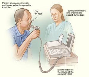
Spirometry: This test assesses how well the lungs are working by measuring air volume. Measurements are taken for respiratory volume, tidal volume, expiratory reserve volume, residual volume, and respiratory capacity. All of these factors play a role in the amount of air we breathe in, keep in our lungs after expiration, and breathe out (Carter & Rutherford, 2020). Fig. 8.83 demonstrates how the procedure is completed as the patient breathes into the machine.
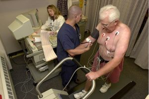
Stress test: Also known as an exercise stress test, this procedure shows how well a patient’s heart works during physical activity (Mayo Clinic, 2022b). It usually involves having the patient walk on a treadmill, as seen in Fig. 8.84, or ride a stationary bike. While the patient is moving, heart rhythm, blood pressure, and breathing are monitored. This test is typically ordered when a patient has a history of cardiac illness or concerns.
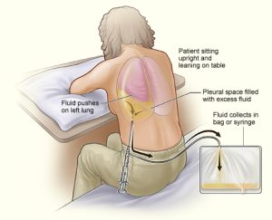
Thoracentesis: This procedure drains fluid from the space between the lungs and the wall of the chest (the pleural space), as shown in Fig. 8.85. A hollow needle or plastic tube is inserted through the chest wall to remove fluid (Canadian Cancer Society, 2022b). This alleviates breathing difficulty and can help diagnose lung issues because a fluid sample is usually sent to the lab for analysis (Carter & Rutherford, 2020). Fig. 8.86 shows a chest X-ray of a patient who requires a thoracentesis for pleural effusion, a condition in which there is fluid build-up in the pleural space, as shown by the white area where the lung should be.

Exercises
Attribution
Unless otherwise indicated, material on this page has been adapted from the following resource:
Betts, J. G., Young, K. A., Wise, J. A., Johnson, E., Poe, B., Kruse, D. H., Korol, O., Johnson, J. E., Womble, M., & DeSaix, P. (2013). Anatomy and physiology. OpenStax. https://openstax.org/details/books/anatomy-and-physiology licensed under CC BY 4.0
References
Canadian Cancer Society. (2022a). Paracentesis. https://cancer.ca/en/treatments/tests-and-procedures/paracentesis
Canadian Cancer Society. (2022b). Thoracentesis. https://cancer.ca/en/treatments/tests-and-procedures/thoracentesis
Cancer Research UK. (2022). Gastroscopy. https://www.cancerresearchuk.org/about-cancer/cancer-in-general/tests/gastroscopy
Carter, K., & Rutherford, M. (2020). Building a medical terminology foundation. eCampus Ontario. https://ecampusontario.pressbooks.pub/medicalterminology/ licensed under CC BY 4.0
Doyle, G. R., & McCutcheon, J. A. (2020). Clinical procedures for safer patient care. BCcampus Open Education. https://opentextbc.ca/clinicalskills/ licensed under CC BY 4.0
Johns Hopkins. (2022). Bronchoscopy. https://www.hopkinsmedicine.org/health/treatment-tests-and-therapies/bronchoscopy#:~:text=Bronchoscopy%20is%20a%20procedure%20to,)%2C%20and%20into%20the%20airways
Mayo Clinic. (2022a). Pap smear. https://www.mayoclinic.org/tests-procedures/pap-smear/about/pac-20394841
Mayo Clinic. (2022b). Stress test. https://www.mayoclinic.org/tests-procedures/stress-test/about/pac-20385234
National Health Service (NHS). (2020). Cystoscopy. https://www.nhs.uk/conditions/cystoscopy/
National Institute of Diabetes and Digestive and Kidney Diseases (NIDDK). (2017). Colonoscopy. https://www.niddk.nih.gov/health-information/diagnostic-tests/colonoscopy
National Library of Medicine (NLM). (2022). Aspiration. https://medlineplus.gov/ency/article/002216.htm#:~:text=Aspiration%20means%20to%20draw%20in,body%20fluids%2C%20or%20bone%20fragments
Nelson, A. (2022). What is phlebotomy? WebMD. https://www.webmd.com/a-to-z-guides/what-is-phlebotomy
Image Credits (images are listed in order of appearance)
Tripler Army Medical Center to roll out Nurse Advice Line 140508-F-AD344-012 by Staff Sgt. Chris Hubenthal, U.S. Air Force, Public domain
Doctor examining child, Seattle, circa 1970s (20851540008) by Seattle Municipal Archives, CC BY 2.0
Amniocentesis by BruceBlaus, CC BY-SA 4.0
Anterior chest auscultation, child by Secom Bahia, CC BY 2.0
Human liver biopsy sample by National Institute of Diabetes and Digestive and Kidney Diseases (NIDDK), Public domain.
Biopsy in formalin by Ajay Kumar Chaurasiya, CC BY-SA 4.0
Bone Marrow Biopsy Needles by Thirteen Of Clubs, CC BY 2.0
Blausen 0097 BoneMarrowBiopsy by Blausen Medical Communications, Inc., CC BY 3.0
Diagram showing a cystoscopy for a man and a woman CRUK 064 by Cancer Research UK, CC BY-SA 4.0
Cystoscopy-im-20050425 by Michael Reeve, CC BY-SA 3.0
Holter monitor NIH by National Heart, Lung, and Blood Institute (NHLBI), Public domain
Cubeholter by Macro987, Public domain
Blausen 0617 LumbarPuncture by BruceBlaus, CC BY 3.0
Exploració física by Amanda Mills, CC0 1.0
ENTApp by B. Welleschik, CC BY-SA 3.0
Blausen 0004 AbdominalParacentesis by BruceBlaus, CC BY 3.0
Wjem-17-189-g001 by Samuel L. Burleson and Henry E. Wang, CC BY 4.0
Spirometry NIH by National Heart, Lung, and Blood Institute (NHLBI), Public domain
Stress test by BlueOctane, Public domain
Thoracentesis by National Heart, Lung, and Blood Institute (NHLBI), Public domain

.jpg)