
6.8 Urinary System
Overview and Functions
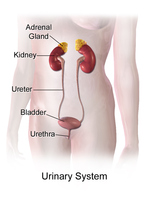
The urinary system (Fig. 6.21) is responsible for cleansing the blood and removing wastes from the body. However, it has other equally important functions, including regulating pH, blood pressure, and the concentration of solutes in the blood; producing erythropoietin (EPO) to stimulate red blood cell production; the final synthesis step of vitamin D production; and producing the active form of vitamin D. The urinary system, controlled by the nervous system, also stores urine until a convenient time for disposal, then provides the structures for transporting liquid waste from the body.
(CrashCourse, 2015)
Components of the Urinary System
Kidneys: The two kidneys are complex organs that perform very complicated and necessary functions that are required for survival. If the kidneys fail, there are devastating effects on the body’s homeostasis. Individuals will experience weakness, lethargy, shortness of breath, anemia, widespread edema, metabolic acidosis, rising potassium levels, heart arrhythmias, and many more symptoms. Nephrons are the functional units of the kidney because they cleanse the blood and balance the components within it (Carter & Rutherford, 2020). They take a simple filtrate of the blood and modify it into urine. Approximately 1 to 2 L of urine are produced and excreted from the body every day.
Ureters: In each kidney, the renal pelvis narrows to become the ureter. As urine passes through the ureter, it does not passively drain into the urinary bladder, but rather is propelled by waves of muscle movement. Each ureter is approximately 30 cm long.
Urinary bladder: The bladder collects urine from both ureters and holds it until it is released from the body via the urethra.
Urethra: This structure transports urine from the bladder to outside the body. The urethra is the only urologic organ that is different between males and females. In women, the urethra is shorter, usually about 4 cm long, whereas in males, it is closer to 20 cm in length. Because of the shorter length of the female urethra, there is less of a barrier to fecal bacteria, so women experience more urinary tract infections (UTIs).
Combining Forms
Table 6.7. Combining Forms
| COMBINING FORM | MEANING | EXAMPLE OF USE IN MEDICAL TERMS |
|---|---|---|
| cyst/o | urinary bladder | cystitis |
| nephr/o | kidney | nephrectomy |
| pyel/o | renal pelvis | pyeloplasty |
| ren/o | kidney | renal |
| ur/o | urine, urinary tract | urologist |
| ureter/o | ureter | ureteroscopy |
| urethr/o | urethra | urethrotomy |
| vesic/o | urinary bladder | vesicostomy |
Common Pathologies
Albuminuria: The presence of protein, or albumin, in the urine (Carter & Rutherford, 2020).
Anuria: No urine production.
Cystitis: This inflammation of the urinary bladder is often caused by an infection (Carter & Rutherford, 2020).
Dysuria: Painful urination (Carter & Rutherford, 2020).
Glomerulonephritis: This condition is acute or chronic nephritis that involves inflammation of the kidney’s filters (glomeruli) (Carter & Rutherford, 2020). The result is that the kidneys lose their ability to remove wastes from the blood to make urine. It has various causes, and if left untreated, it can lead to kidney failure.
Glycosuria: The presence of glucose in the urine.
Hematuria: The presence of blood in the urine (Chabner, 2018).
Hydronephrosis: This is a condition in which the kidneys begin to swell because of urine retention. It is often caused by kidney stones and blood clots (Carter & Rutherford, 2020).
Incontinence: The loss of the ability to control micturition (releasing urine from the bladder).
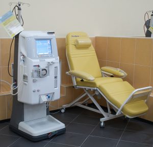
Kidney failure: This occurs when the kidneys lose all, or some, of the ability to produce urine and filter the blood. Symptoms include weakness, lethargy, shortness of breath, edema, anemia, metabolic acidosis, heart arrhythmias, uremia, loss of appetite, and/or an increase or decrease in urination. The treatment for kidney failure is dialysis (Carter & Rutherford, 2020). Hemodialysis is an ongoing treatment in which the individual goes into the hospital regularly to have their blood processed through a dialysis machine (Fig. 6.22), which removes the waste products and returns the blood to the person’s body (Carter & Rutherford, 2020).
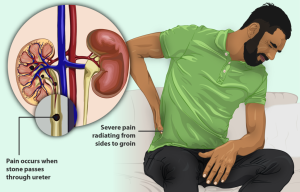
Kidney stones: These hard deposits, typically made up of minerals and salts, form inside the kidney and can cause abdominal pain and discomfort (Fig. 6.23). This condition is also referred to as nephrolithiasis (Carter & Rutherford, 2020).
Oliguria: This condition is characterized by below-normal output of urine of approximately 300 to 500 mL per day. It can be caused by dehydration, blood loss, diarrhea, cardiogenic shock, kidney disease, or an enlarged prostate.
Polycystic kidney disease (PKD): This is a genetic disease in which cysts grow inside the kidneys. The kidneys then enlarge and damage occurs to the filtering structures of the kidneys (Carter & Rutherford, 2020).
Polyuria: This condition is characterized by excessive urine production of more than 2.5 L a day. It can be caused by diabetes mellitus, excessive intake of caffeine or alcohol, kidney disease, drugs similar to diuretics, sickle cell anemia, and excessive water intake.
Renal cell carcinoma: This is a cancer that occurs in the kidney tubes where urine is produced or collected. It is one of the most common cancers that occurs within the kidneys (Carter & Rutherford, 2020).
Uremia: The retention of urea and uric acid in the blood.
Urinary tract infection (UTI): This infection is caused by bacteria or sometimes fungi. The exact type of bacterial growth is determined by collecting urine for culture and sensitivity (C&S) testing (Carter & Rutherford, 2020). Results from the C&S test will determine the best treatment options for the individual.
Exercise
Attribution
Unless otherwise indicated, material on this page has been adapted from the following resource:
Betts, J. G., Young, K. A., Wise, J. A., Johnson, E., Poe, B., Kruse, D. H., Korol, O., Johnson, J. E., Womble, M., & DeSaix, P. (2013). Anatomy and physiology. OpenStax. https://openstax.org/details/books/anatomy-and-physiology licensed under CC BY 4.0
References
Carter, K., & Rutherford, M. (2020). Building a medical terminology foundation. eCampusOntario. https://ecampusontario.pressbooks.pub/medicalterminology/ licensed under CC BY 4.0
Chabner, D. E. (2018). Medical terminology: A short course (8th ed.). Saunders/Elsevier.
CrashCourse. (2015, October 12). Urinary system, part I: Crash Course A&P #38 [Video]. YouTube. https://www.youtube.com/watch?v=l128tW1H5a8&list=PL8dPuuaLjXtOAKed_MxxWBNaPno5h3Zs8&index=40
Image Credits (images are listed in order of appearance)
Urinary System (Female) by BruceBlaus, CC BY-SA 4.0
Hemodialysis machine INNOVA by Виталий Поспелов, CC BY 3.0
A man with Kidney Stones by myUpchar, CC BY-SA 4.0
Inflammation of the urinary bladder
Removal of a kidney
Surgical correction of the renal pelvis
Pertaining to the kidneys
A specialist in the study of the urinary tract
The process of visually examining the ureters (upper urinary tract)
An incision into the urethra
An incision into the urinary bladder

.png)