
6.12 Musculoskeletal System
Overview and Functions
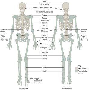
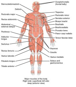
The musculoskeletal system consists of all the bones, muscles, joints, tendons, and cartilage found in the human body (Figs. 6.39 and 6.40). The purpose of this system is to support the body, facilitate movement, and protect the internal organs. Bones are also vital in the production of red blood cells. Some resources show the muscular and skeletal systems as separate; however, for the purposes of this book, they are combined in order to provide a basic overview of their components, functions, and pathologies.
(CrashCourse, 2015a)
(CrashCourse, 2015b)
Components of the Musculoskeletal System
The skeleton is subdivided into two major components:
- Axial skeleton: The axial skeleton forms the vertical, central axis of the body and includes all the bones of the head, neck, chest, and back. It protects the brain, spinal cord, heart, and lungs. It also serves as the attachment site for muscles that move the head, neck, and back, and for muscles that act across the shoulder and hip joints to move their corresponding limbs. There are 80 bones in the axial skeleton.
- Appendicular skeleton: The appendicular skeleton includes all the bones of the upper and lower limbs, plus the bones that attach each limb to the axial skeleton. There are 126 bones in the appendicular skeleton.
Bones: There are 206 bones that compose the adult skeleton. Bones are also known as osseous tissue and are hard, dense connective tissue. The purpose of bones is to assist with movement, protect the organs, and produce red blood cells.
Muscle: This is one of the four primary tissue types of the body. The body contains three kinds of muscle tissue: skeletal muscle, cardiac muscle, and smooth muscle.
Joints: These are also known as articulation and are any place where adjacent bones or bone and cartilage come together to form a connection.
Tendons: These dense, fibrous connective tissues anchor muscle to bone.
Cartilage: This elastic connective tissue is found at the ends of bones as well as in other locations such as the tip of the nose.
Ligaments: These tough, elastic connective tissues connect bone to bone.
Combining Forms
Table 6.11. Combining Forms
| COMBINING FORM | MEANING | EXAMPLE OF USE IN MEDICAL TERMS |
|---|---|---|
| arthr/o | joint | arthritis |
| cervic/o | neck | cervical |
| chondr/o | cartilage | chondrocytes |
| coccyg/o | coccyx, tailbone | coccygeal |
| cost/o | rib | costectomy |
| crani/o | skull | craniotomy |
| ligament/o | ligament | ligamentitis |
| lumb/o | loin, waist | lumbar |
| muscul/o | muscle | muscular |
| my/o | muscle | myectomy |
| myos/o | muscle | myositis |
| myel/o | bone marrow | myeloma |
| odont/o | tooth | orthodontist |
| oste/o | bone | osteomyelitis |
| pelv/o | pelvis, hip bone | pelvic |
| sacr/o | sacrum | sacroiliitis |
| spin/o | spine, backbone | spinal stenoisis |
| spondyl/o | vertebra | spondylosis |
| ten/o | tendon | tenotomy |
| tendin/o | tendon | tendinopathy |
| vertebr/o | vertebra | vertebral |
Common Pathologies
Ankylosing scoliosis: This is a lateral curvature and twist of the spine. It is most common among girls and typically gets worse during adolescent growth spurts. Many people who have this pathology do not require treatment, but some must wear a back brace or, in rare instances, may require surgery.
Arthritis: By definition, arthritis means “inflammation (-itis) of the joint (arthr/o).” Individuals with arthritis often present with joint pain, redness, and swelling. There is no cure for arthritis, and it is treated through exercise, medications, and, in some cases, joint replacement.
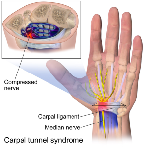
Carpal tunnel syndrome: This pathology is common among those with occupations requiring repetitive movements of the hand, such as office workers and cashiers. It is characterized by pain or numbness accompanied by muscle weakness in the area of the wrist and hand where the median nerve becomes compressed (Fig. 6.41).
Gout: This condition occurs when uric acid builds up in a joint. Symptoms of gout include swelling, pain, and redness in the affected joint. Gout can flare up, then dissipate and reoccur at a later date. It can affect any joint, but most often occurs in the toes.
Kyphosis: This is a forward curvature of the spine in the thoracic (upper back) region.
Lordosis: This is an excessive inward (anterior) curvature of the spine in the lumbar region and is often associated with obesity or the late stages of pregnancy. It is sometimes called swayback.
Muscular dystrophy: This condition is a progressive weakening of the skeletal muscles. It is an inherited disorder and mostly affects males. Symptoms usually start with balance issues and then progress to inability to walk. Eventually, muscular dystrophy causes respiratory failure and death.
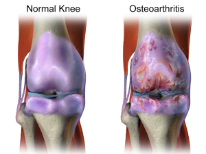
Osteoarthritis: This is the most common form of arthritis and involves the breakdown of cartilage and bone over time (Fig. 6.42). The most common symptoms are pain and stiffness that progressively worsen.
Osteomyelitis: This is an infection within the bone that is caused by staphylococcus bacteria. The bacteria can travel through the bloodstream to the bone or gain access via a wound or surgery (Carter & Rutherford, 2020).
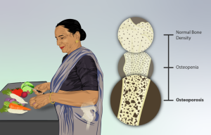
Osteoporosis: This pathology is characterized by progressive bone loss that causes bones to become weak and thin over time (Fig. 6.43). There can be an increased chance of fractures because the bones are weakened. The likelihood of developing osteoporosis increases with age, and the condition is more common in females.
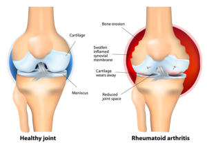
Rheumatoid arthritis (RA): This autoimmune disease presents with inflammation to the joint tissues of the hands, wrists, and knees. Symptoms include debilitating pain and swelling in the affected area (Fig. 6.44).
Exercise
Attribution
Unless otherwise indicated, material on this page has been adapted from the following resource:
Betts, J. G., Young, K. A., Wise, J. A., Johnson, E., Poe, B., Kruse, D. H., Korol, O., Johnson, J. E., Womble, M., & DeSaix, P. (2013). Anatomy and physiology. OpenStax. https://openstax.org/details/books/anatomy-and-physiology licensed under CC BY 4.0
References
Carter, K., & Rutherford, M. (2020). Building a medical terminology foundation. eCampusOntario. https://ecampusontario.pressbooks.pub/medicalterminology/ licensed under CC BY 4.0
CrashCourse. (2015a, May 18). The skeletal system: Crash Course A&P #19 [Video]. YouTube. https://www.youtube.com/watch?v=rDGqkMHPDqE&list=PL8dPuuaLjXtOAKed_MxxWBNaPno5h3Zs8&index=20
CrashCourse. (2015b, June 15). Muscles, part 2 – Organismal level: Crash Course A&P #22 [Video]. YouTube. https://www.youtube.com/watch?v=I80Xx7pA9hQ&list=PL8dPuuaLjXtOAKed_MxxWBNaPno5h3Zs8&index=23
Image Credits (images are listed in order of appearance)
701 Axial Skeleton-01 by OpenStax, CC BY 3.0
1105 Anterior and Posterior Views of Muscles by OpenStax, CC BY-SA 4.0
Carpal Tunnel Syndrome by BruceBlaus, CC BY 3.0
Osteoarthritis by BruceBlaus, CC BY-SA 4.0
Depiction of an Osteoporosis patient by myUpchar, CC BY-SA 4.0
Rheumatoid-Arthritis by National Library of Medicine, Public domain
Inflammation of the joints
Pertaining to the cervix
Cells in cartilage; the only cells found in healthy cartilage
Pertaining to the coccyx
Removal of all or part of a rib
Cutting into the skull
Inflammation of a ligament
Pertaining to the loins or waist
Pertaining to muscle
Removal of part or all of a muscle
Inflammation of muscle
A malignant tumour in the bone marrow
A dentist who specializes in straightening teeth and correcting bites
Inflammation of the bone and muscle
Pertaining to the pelvis
Inflammation of the sacrum and ileum (around the joint)
Narrowing of the space within the spine; can cause compression of the spinal nerves
A degenerative condition of the spine cause by ongoing wear and tear
An incision into a tendon
Disease condition of a tendon
Pertaining to the vertebra
