
5.5 Divisions of the Spine
The spine is also known as the spinal column or vertebral column. It consists of vertebrae (singular = vertebra) separated by intervertebral discs. Together, the vertebrae and intervertebral discs form the spine, which is a flexible column that supports the head, neck, and body, and allows for movement. It also protects the spinal cord, which passes down the spine through openings in the vertebrae.
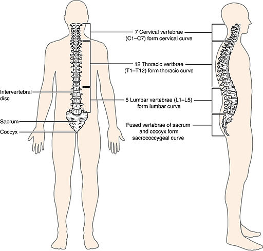
The spine originally has a total of 33 vertebrae; however, this number decreases with age to 24 vertebrae, plus the sacrum and coccyx. It is divided into five regions, with the vertebrae in each area named for that region and numbered in increasing order from top to bottom. Fig. 5.20 and Table 5.4 provide a breakdown of the sections of the spine.
The neck has seven cervical vertebrae, each designated C1-C7. Below these are the 12 thoracic vertebrae, designated T1–T12. The lower back contains the lumbar vertebrae, numbered L1–L5. The sacrum is formed by the fusion of five sacral vertebrae. Similarly, the coccyx, or tailbone, results from the fusion of four small coccygeal vertebrae. The sacral and coccygeal fusions, which result in the decrease in total number of vertebrae, do not begin until approximately age 20, and the fusions are not completed until a person is close to middle age.
Table. 5.4 Divisions of the Spine
| DIVISION | BONES | ABBREVIATION |
| cervical | 7 bones | C1-C7 |
| thoracic | 12 bones | T1-T12 |
| lumbar | 5 bones | L1-L5 |
| sacral | 5 fused bones | |
| coccygeal | 4 fused bones |
Did you know?
Interestingly, almost all mammals have seven cervical vertebrae, regardless of their body size (Carter & Rutherford, 2020). This means that the size of the cervical vertebrae vary widely. They range from the tiny cervical vertebrae of a bird to the large vertebrae in the neck of a giraffe. In a full-grown giraffe, each cervical vertebra measures 28 centimetres (11 inches) tall (Carter & Rutherford, 2020).
The adult spine does not form a straight line, but instead has four curvatures along its length. The purpose of these curves is to increase the spine’s strength, flexibility, and shock-absorbing ability. For example, when you are carrying a heavy backpack, the spine becomes more curved to accommodate the extra weight, then springs back when the weight is removed.
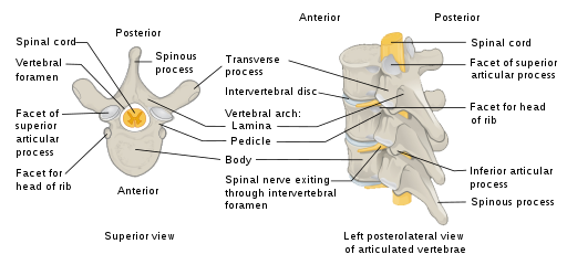
In the different regions of the spine, the vertebrae vary in size and shape, but they all follow a similar structural pattern. A typical vertebra consists of a body, a vertebral arch, and seven processes. The body is the front portion of each vertebra and supports the body’s weight. The vertebral arch forms the back portion of each vertebra. The large opening between the vertebral arch and body contains the spinal cord. The seven processes are outgrowths that arise from the vertebral arch. They serve various functions, including attachments for muscles and ribs. The structure and components of the vertebrae can be seen in Fig. 5.21.
A lumbar puncture is a common diagnostic test in which a needle is inserted into the lumbar region of the spinal column through the dura mater and arachnoid membrane into the subarachnoid space so that fluid can be withdrawn for testing. Because the spinal cord does not extend through the lower lumbar region of the spine, a needle can be inserted through the dura and arachnoid layers to withdraw cerebrospinal fluid and avoids the risk of damaging the central tissue of the spinal cord. This procedure is also called a spinal tap, and the common abbreviation is LP. Fig. 5.22 shows a spinal tap being performed, and Fig. 5.23 demonstrates how a sample of fluid is taken for analysis.
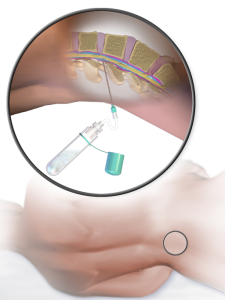
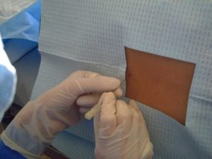
Table. 5.5. Combining Form Review
| COMBINING FORM | MEANING |
| cervic/o | neck |
| chondr/o | cartilage |
| coccyg/o | coccyx, tailbone |
| lamin/o | part of a vertebra/backbone |
| lumb/o | loin, waist |
| myel/o | spinal cord |
| pelv/o | pelvis |
| sacr/o | sacrum |
| spin/o | spine |
| vertebr/o | vertebra |
(Carter & Rutherford, 2020)
Exercises
Attribution
Unless otherwise indicated, material on this page has been adapted from the following resource:
Betts, J. G., Young, K. A., Wise, J. A., Johnson, E., Poe, B., Kruse, D. H., Korol, O., Johnson, J. E., Womble, M., & DeSaix, P. (2013). Anatomy and physiology. OpenStax. https://openstax.org/details/books/anatomy-and-physiology licensed under CC BY 4.0
References
Carter, K., & Rutherford, M. (2020). Building a medical terminology foundation. eCampusOntario. https://ecampusontario.pressbooks.pub/medicalterminology/ licensed under CC BY 4.0
Image Credits (images are listed in order of appearance)
715 Vertebral Column by OpenStax, CC BY 3.0
Vertebra en by Jmarchn, CC BY-SA 3.0
Puncion lumbar by Dragondefuego1976, CC BY-SA 4.0
Spinal Tap by Medical Gallery of Blausen Medical 2014, BruceBlaus, CC BY 3.0
