
5.4 Body Cavities and the Abdominal Regions and Quadrants
There are several cavities within the body. Each cavity contains a number of organs and plays a specific role in the functioning of the body. We will first examine the body cavities and the organs contained within them. Next, we will look at the nine abdominal regions and four quadrants and relate this topic to patient care.
Body Cavities and Serous Membranes
The dorsal (posterior) cavity and the ventral (anterior) cavity are the largest body compartments. Fig 5.17 shows the body divided into the many body cavities. These cavities contain and protect delicate internal organs, and the ventral cavity allows for significant changes in the size and shape of the organs as they perform their functions. For example, the lungs, heart, stomach, and intestines can expand and contract without distorting other tissues or disrupting the activity of nearby organs.
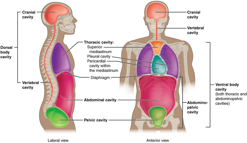
The posterior (dorsal) and anterior (ventral) cavities are each subdivided into smaller cavities:
The posterior (dorsal) cavity has two main subdivisions:
- The cranial cavity houses the brain and pineal and pituitary glands.
- It is protected by the bones of the skull and the cerebrospinal fluid.
- The spinal (vertebral) cavity encloses the spinal cord.
- It is protected by the spinal column and the cerebrospinal fluid.
The anterior (ventral) cavity has two main subdivisions:
- The thoracic cavity is the more superior subdivision of the anterior cavity and is enclosed by the rib cage.
- The thoracic cavity contains the heart, lungs, trachea, and bronchial tubes.
- The mediastinum is located between the lungs and contains the heart, trachea, esophagus, and bronchial tubes.
- The pericardial cavity is within the mediastinum and is formed from the pericardium, which is a double membrane that surrounds the heart.
- The pleural cavity is a smaller cavity within the thoracic cavity. The lungs are protected by a double membrane called the pleura, and the space between the pleura is the pleura cavity.
- The diaphragm is a muscle that forms the floor of the thoracic cavity and separates it from the more inferior abdominopelvic cavity.
- The abdominopelvic cavity is the largest cavity in the body.
- No membrane physically divides the abdominopelvic cavity.
- The abdominopelvic cavity houses both the abdominal cavity and the pelvic cavity.
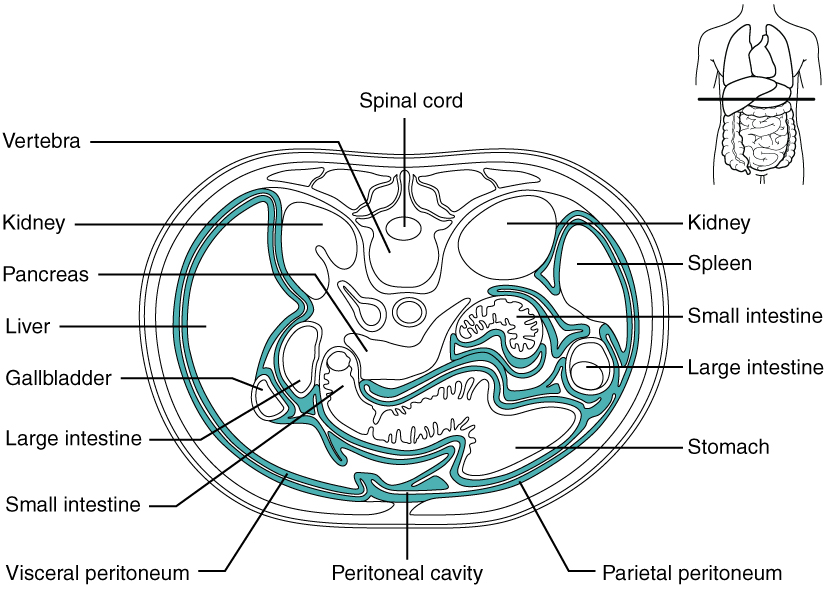
- The abdominal cavity houses the digestive organs (stomach, liver, gallbladder, and small and large intestines).
- The organs in the abdominal cavity are surrounded by a double membrane called the peritoneum. The space between this double membrane is called the peritoneal cavity. This membrane adds protection and supports and reduces friction between the abdominal and pelvic organs. Fig. 5.18 shows the peritoneum (highlighted in green) and the peritoneal cavity in a transverse (axial) view.
- The pelvic cavity is inferior to the abdominal cavity. It contains the male or female reproductive organs, urinary system organs (urinary bladder, ureters, urethra), rectum, and anus.
Key Concepts
Sometimes fluid can build up in the different body cavities, causing strain on the body (Mayo Clinic, 2022a). Starting with the most superior body cavity, the cranial cavity, hydrocephalus is a condition in which there is an increase in fluid (cerebrospinal fluid) in the cranial cavity. This in turn puts increased pressure on the brain and can cause numerous issues, including headaches, visual disturbances, and balance problems, among others. This condition usually needs to be treated both medically and surgically (Mayo Clinic, 2022a).
The thoracic cavity has two main cavities where fluid can build up—the pleural cavity and the pericardial cavity (Cleveland Clinic, 2018). A collection of fluid in the pleural cavity is called pleural effusion. Fluid build-up in the pericardial cavity is called pericardial effusion (Mayo Clinic, 2022b). In both these conditions, when there is a build-up of fluid within their double membranes, patients may present with angina and dyspnea (Cleveland Clinic, 2018; Mayo Clinic, 2022b).
Fluid build-up in the peritoneal cavity is called ascites (Cleveland Clinic, 2021). Ascites can happen for a number of reasons, most commonly cirrhosis of the liver. Patients usually present with dyspnea and swelling in the abdomen and ankles (Cleveland Clinic, 2021).
Abdominal Regions and Quadrants
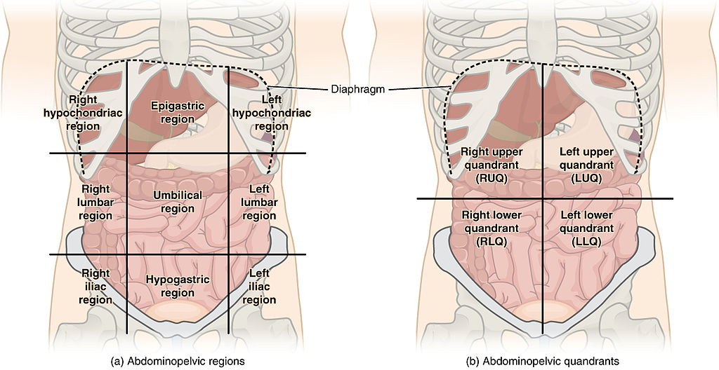
To promote clear communication, for instance, about the location of a patient’s abdominal pain or a suspicious mass, healthcare providers typically divide up the abdominal cavity into either nine regions or four quadrants. Fig. 5.19 shows both (a) the nine abdominopelvic regions and (b) the four quadrants. It is important to be aware of the major organs in each region or quadrant. For example, the stomach is in the left upper quadrant (LUQ), and the appendix sits in the right lower quadrant (RLQ).
Key Concept
The nine abdominopelvic regions and the four quadrants are often used when describing a mass or pain. It makes communication among healthcare provides more clear. For example, if a patient has a mass in the left lower quadrant it will be written as “patient has a mass in the LLQ.” This way, when another healthcare provider goes to reassess the patient, they know exactly what area of the abdomen they need to look at.
Table 5.3. Combining Form Review
| COMBINING FORM | MEANING |
| abdomin/o | abdomen |
| anter/o | front |
| cephal/o | head |
| crani/o | skull |
| hydr/o | water |
| lapar/o | abdomen |
| mediastin/o | mediastinum |
| myel/o | spinal cord |
| pelv/o | pelvis |
| peritone/o | peritoneum |
| phren/o | diaphragm |
| pleur/o | pleura |
| poster/o | back, behind |
| spin/o | spine |
| spondyl/o | backbone, vertebra |
| thorac/o | chest |
(Carter & Rutherford, 2020)
Exercise
Exercises
Attribution
Unless otherwise indicated, material on this page has been adapted from the following resource:
Betts, J. G., Young, K. A., Wise, J. A., Johnson, E., Poe, B., Kruse, D. H., Korol, O., Johnson, J. E., Womble, M., & DeSaix, P. (2013). Anatomy and physiology. OpenStax. https://openstax.org/details/books/anatomy-and-physiology licensed under CC BY 4.0
References
Carter, K., & Rutherford, M. (2020). Building a medical terminology foundation. eCampusOntario. https://ecampusontario.pressbooks.pub/medicalterminology/ licensed under CC BY 4.0
Cleveland Clinic. (2021). Ascites. https://my.clevelandclinic.org/health/diseases/14792-ascites
Cleveland Clinic. (2018). Pleural effusion causes, signs & treatment. https://my.clevelandclinic.org/health/diseases/17373-pleural-effusion-causes-signs–treatment#:~:text=Pleural%20effusion%2C%20sometimes%20referred%20to,to%20lubricate%20and%20facilitate%20breathing
Mayo Clinic. (2022a). Hydrocephalus. https://www.mayoclinic.org/diseases-conditions/hydrocephalus/symptoms-causes/syc-20373604#:~:text=Overview,the%20brain%20and%20spinal%20column
Mayo Clinic. (2022b). Pericardial effusion. https://www.mayoclinic.org/diseases-conditions/pericardial-effusion/symptoms-causes/syc-20353720#:~:text=Pericardial%20effusion%20(per%2De%2D,a%20thin%20layer%20of%20fluid
Image Credits (images are listed in order of appearance)
Body cavities by OpenStax, CC BY 3.0
2403 The PeritoneumN by OpenStax, CC BY 3.0
Abdominal Quadrant Regions by OpenStax, CC BY 3.0
Chest pain
Pain or difficulty breathing
