
6.2 Cardiovascular System
Overview and Functions
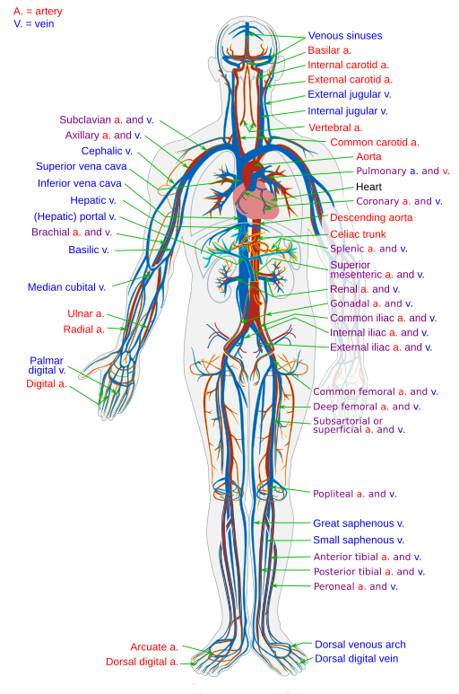
The cardiovascular system (Fig. 6.1) uses blood to deliver nutrients and remove wastes from the trillions of cells in the human body. The heart, which is the primary organ in this system, pumps blood throughout the body via a network of blood vessels. These three components—blood, blood vessels, and the heart—make up this complex system. Figure 6.1 illustrates the cardiovascular system, and the video provided explains the intricate function of the heart within this system.
(CrashCourse, 2015)
Components of the Cardiovascular System
Heart: The human heart is located in the thoracic cavity, between the lungs in the space referred to as the mediastinum. The best way to describe the heart is a pump. Its contractions create pressure that sends blood into the major vessels, including the aorta. From there, the blood is distributed to the rest of the body.
Blood: Blood is composed of many different compounds and can be analyzed using lab tests to determine the various levels of these compounds present in the body. The main components of blood are the following:
- Plasma: Consists of water, proteins, nutrients, and hormones
- Hematocrit: Consists of red blood cells
- Buffy coat: Consists of white blood cells and platelets
The primary function of blood is to deliver oxygen and nutrients to the body’s cells and to remove wastes from those same cells. Blood also has other functions, including defence, distribution of heat, and maintenance of homeostasis.
Blood vessels: Blood vessels transport blood throughout the body. They provide the physical site where gases, nutrients, and other substances are exchanged with body cells. An artery is a blood vessel that carries blood away from the heart. It branches into smaller vessels called arterioles, and they further branch into tiny capillaries, where nutrients and wastes are exchanged. The capillaries then combine to form venules, which are small blood vessels that carry blood to a vein, a larger blood vessel that returns blood to the heart.
Combining Forms
Table 6.1. Combining Forms
| COMBINING FORM | MEANING | EXAMPLE OF USE IN MEDICAL TERMS |
|---|---|---|
| angi/o | blood vessel | angioplasty |
| aort/o | aorta | aortic |
| arteri/o | artery | arteriosclerosis |
| arteriol/o | arteriole | arteriolitis |
| cardi/o | heart | cardiomegaly |
| coron/o | heart | coronary |
| hem/o | blood | hemoglobin |
| hemat/o | blood | hematoma |
| mediastin/o | mediastinum | mediastinotomy |
| phleb/o | vein | phlebotomy |
| vas/o | blood vessel | vasoconstriction |
| ven/o | vein | intravenous |
| venul/o | venule | venulectasia |
Common Pathologies
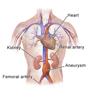
Aneurysm: An aneurysm (Fig. 6.2) is the ballooning of a blood vessel caused by weakening of the wall of the vessel. It can occur in various vessels throughout the body, including the brain and abdomen. An aneurysm can be a life-threatening emergency, especially if the aneurysm bursts, which can lead to the person bleeding out internally.
Angina: Angina is characterized by chest pain and is caused by poor blood flow to the heart. Often it can be treated with nitroglycerin, a vasodilator medication that increases blood flow back to and through the heart, and rest.
Arteriosclerosis: This condition is defined as hardening of the arteries. Advanced age and lifestyle factors such as smoking, high blood sugar, infection, and high cholesterol are major risk factors for arteriosclerosis, which progresses over time. This pathology begins with some form of injury to the walls of the arteries, which results in inflammation to the area. The inflammation then spreads throughout the walls of the artery, causing weakness and scarring. Over time, the scarring results in the arteries becoming stiff or “hardened” (Ernstmeyer & Christman, 2020).
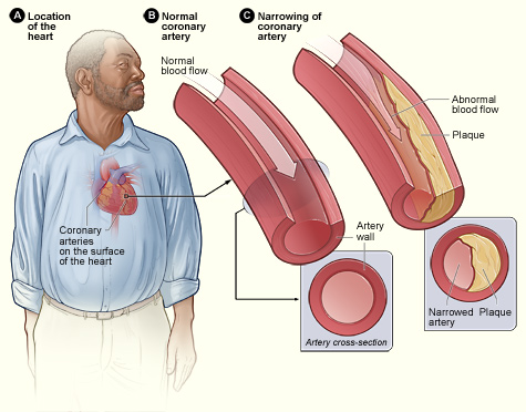
Atherosclerosis: This is a type of arteriosclerosis and is characterized by a build-up of plaque along the walls of the arteries (Fig. 6.3). The plaque is a fatty material made up of cholesterol, white blood cells, connective tissue, and smooth muscle cells. The plaque build-up obstructs blood flow and decreases the flexibility of the arteries.
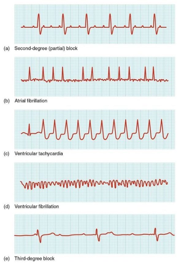
Arrhythmia: Any deviation from the normal pattern of impulses and contractions within the heart is considered an arrhythmia. It can also be any form of fibrillation, which is uncoordinated beating of the heart. An electrocardiogram (Fig. 6.4) provides a record of the electrical activity of the heart and can help diagnose this condition.
Deep vein thrombosis (DVT): A deep vein thrombosis is a blood clot that forms in one of the larger blood vessels, often in the calf of the leg. It is more likely to occur after a long plane flight or period of immobility. The clot can result in a pulmonary embolism, which is a blood clot that travels to the lungs.
Heart failure: In this condition, the heart is unable to pump enough blood to supply the body. Right-sided heart failure occurs when the heart cannot pump enough blood to the lungs, where it picks up oxygen (Ernstmeyer & Christman, 2020). Left-sided heart failure, on the other hand, occurs when the heart cannot pump enough oxygen-filled blood to the rest of the body. There is no cure for this pathology; however, it can be treated with lifestyle changes and medications. Individuals with heart failure often exhibit peripheral edema (swelling of the hands and feet) and shortness of breath because of fluid overload in the lungs (Ernstmeyer & Christman, 2020).
Hyperlipidemia: High blood lipid levels or high circulating levels of lipids in the blood characterize this pathology. Cholesterol is a fat, also called a lipid, and it is necessary for our bodies to work correctly (Ernstmeyer & Christman, 2020). There are different types of cholesterol, and they include the following:
- High-density lipoprotein (HDL) cholesterol: This is considered a “good” cholesterol because it promotes the excretion of cholesterol (Ernstmeyer & Christman, 2020).
- Low-density lipoprotein (LDL) cholesterol: This is often called “bad” cholesterol because it stores cholesterol in the bloodstream, which contributes to atherosclerosis (Ernstmeyer & Christman, 2020). Too much LDL cholesterol can increase the risk for a stroke, peripheral vascular disease, and various forms of heart disease.
- Total cholesterol: This is all the cholesterols combined.
Hypertension (HTN): Hypertension is abnormally high blood pressure. The previous standard for this diagnosis was 140/90 mmHg, but recent guidelines have come out that state hypertension should begin to be treated at 130/80 mmHg (Ernstmeyer & Christman, 2020). This condition is treated with lifestyle changes and medications.
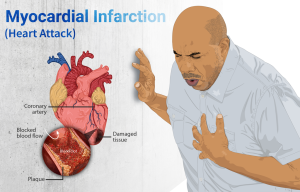
Myocardial infarction (MI): This is the medical term for what is commonly referred to as a heart attack (Fig. 6.5). It results from lack of blood flow and oxygen to a part of the heart. The lack of blood flow to that area (ischemia) then results in the death of the tissues (necrosis). Symptoms can vary but often present with chest pain that radiates to the back or shoulder, as well as paleness and diaphoresis (sweating).
Shock: There are many different types of shock; however, all of them are characterized by the inability of the body to get oxygen to the tissues and insufficient return of blood to the heart. Symptoms can vary and usually include paleness, rapid and weak pulse, and shortness of breath. Some types of shock include the following:
- Cardiogenic shock: Occurs when the heart cannot maintain cardiac output
- Anaphylactic shock: Results from a severe allergic reaction and massive vasodilation
- Circulatory shock: Occurs when the body cannot supply enough blood flow for adequate oxygenation of the tissues
- Hypovolemic shock: Excessive loss of blood volume caused by dehydration or hemorrhage
- Septic shock: Results from a severe infection and inflammation
Exercise
Attribution
Unless otherwise indicated, material on this page has been adapted from the following resource:
Betts, J. G., Young, K. A., Wise, J. A., Johnson, E., Poe, B., Kruse, D. H., Korol, O., Johnson, J. E., Womble, M., & DeSaix, P. (2013). Anatomy and physiology. OpenStax. https://openstax.org/details/books/anatomy-and-physiology licensed under CC BY 4.0
References
CrashCourse. (2015, July 6). The heart, part 1 – Under pressure: Crash Course A&P #25 [Video]. YouTube. https://www.youtube.com/watch?v=X9ZZ6tcxArI&list=PL8dPuuaLjXtOAKed_MxxWBNaPno5h3Zs8&index=26
Ernstmeyer, K., & Christman, E. (Eds.). (2020). Nursing pharmacology. Chippewa Valley Technical College. https://wtcs.pressbooks.pub/pharmacology/ licensed under CC BY 4.0
Image Credits (images are listed in order of appearance)
Circulatory System en by LadyofHats, Public domain
Abdominal Aortic Aneurysm Location by BruceBlaus, CC BY-SA 4.0
Atherosclerosis 2011 by National Heart, Lung, and Blood Institute, Public domain
2024 Cardiac Arrhythmias by OpenStax, CC BY 3.0
Depiction of a person suffering from a heart attack (Myocardial Infarction) by myUpchar, CC BY-SA 4.0
The surgical repair of a blood vessel
Pertaining to the aorta
The hardening of an artery
Inflammation of the arterioles
Enlargement of the heart
Pertaining to the heart
A protein in the blood that carries oxygen
A mass of blood
An incision into the mediastinum
An incision into a vein
Narrowing of a blood vessel
Pertaining to within a vein
A large, palpable type of spider vein that is usually purple

.png)